| Annals of Burns and Fire Disasters - vol. XI - n. 1 - March 1998
ASSESSMENT OF CERTAIN NEUTROPHIL RECEPTORS,
OPSONOPHAGOCYTOSIS AND SOLUBLE INTERCELLULAR ADHESION MOLECULE.1 (ICAM-1) FOLLOWING
THERMAL INJURY
Shehab El-Din S.A., Aref S.E., Salama O.S.
Faculty of Medicine, Mansoura University,
Mansoura, Egypt
SUMMARY. Polymorphnuclear
leukocytes (PMLs) play a key role in host defence, and phagocyte dysfunction has been
associated with increased susceptibility to infections in patients with thermal injury.
Intercellular adhesion molecule-1 (ICAM-1) plays a role in leukocyte accumulation and
extravasation. Flow cytometric analysis (FCM) was used to study PML expression of IgG
Fc-receptor III (Fey R111) as well as the complement receptors CRI (receptor for C3b) and
CR3 (receptor for Obi) in 23 patients with large burns. Analysis of PML complement- and
immunoglobulin-mediated phagocytosis of Candida albicans was performed in parallel
using the phagocytic index. Plasma ICAM-1 was determined using ELISA. This study revealed
a significant increase, with variable degrees, in CRI- and CR3-dependent fluorescence,
complement-mediated phagocytosis of Candida albicans, and plasma ICAM- 1, starting
on day 2 and continuing for about 20 days until normalization. In contrast Fcy
RIII-dependent fluorescence and Ig-mediated phagocytosis were significantly decreased
versus control values. These results demonstrate a significant change in PML opsonin
receptor expression and opsonophagocytosis, documenting systemic activation of PMLs after
large burns. In addition, elevation of plasma ICAM-1 may enhance the harmful effect of
neutrophil activation due to leukocyte accumulation and extravasation following
enclothelial damage in the skin and lung.
Introduction
In spite of marked improvements in
fluid resuscitation, respiratory care techniques, and other intensive care procedures
introduced in the last decades, infection continues to be the leading cause of death in
thermally injured patients.' Loss of the protective skin barrier, nutritional imbalance,
and increased metabolic requirements contribute to the increased susceptibility to
infection. In addition, thermal injury induces profound abnormalities in specific and
unspecific immunity, exposing the patients to considerable risk of infection!
Polymorphnuclear leukocyte (PMLs) are effector cells essential for protection against
bacterial and fungal infection? Immunoglobulins and complement factors serve as opsonins
and facilitate phagocytosis via specific opsonin receptors. The most important opsonin
receptors include Fcy RII and Fcy RIII for IgG, besides CRI and CR3 for the C3 split
products C3b and ON, respectively! The expression of these receptors is modulated by
chemoattractants and cytokines, as well as by injury and various diseases.
Human Fcy receptor III (FCRIII) or CD16-antigen is expressed on neutrophils, natural
killer (NK) lymphocytes, and macrophages. Two genes are coding for this receptor, FcRIII-1
and FcRIII-2. The FcRIII-I mRNA encodes a protein with a short (four amino acids)
cytoplasmic domain, whereas the FcRIII-2 mRNA encodes a protein with a cytoplasmic domain
of 25 amino acids. The FcRIII-I protein is proteolytically split during post-translational
processing and coupled to a phosphatidylinositol (PI) anchor, whereas the FcRIII-2 protein
appears to be a transmembrane protein. Neutrophils appear to express only the Pllinked
form of FcRIll, while NK lymphocytes and macrophages express only the transmembrane form
of FcRIII.
The two distinct receptors for opsonic fragments of C3 on human phagocytic cells that have
been identified designated CRI and CR3. CRI binds C3b with higher affinity than ON and has
been found to be a membrane glycoprotein with an apparent m.w. of 205,000 to 250,000. This
protein mediates the binding of Ob-coated particles and immune complexes to a variety of
cells bearing the receptor, including neutrophils, monocytes, macrophages, B lymphocytes,
a subset of T lymphocytes, and glomerular podocytes.' CR3 binds Obi-coated particles. CR3,
a membrane heterodimer present on human PMLs, monocytes and null cells, consists of two
non-covalently linked polypeptides with m.w. of 155,000 to 170,000 and 94,000."
Leucocyte stimulation with a variety of agents augmented the expression of CRI and CR3.
Cell membrane expression of adhesion molecules is iniportant for cellular interactions,
including interactions during an immune response." In addition, adhesion molecules
can be detected in vivo," and soluble adhesion molecules may then inhibit binding
between membrane-bound adhesion molecules and their ligands. 14 ICAM-1 is expressed on
many different cells and its expression can be induced by IL-I and TNF-a.." Serum
concentrations of ICAM-1 can be increased during immune or inflammatory disorders.
Alterations of PML chemotaxis," phagocytosis," oxidative metabolism," and
intracellular killing" have been demonstrated after thermal injury.
The present study was performed with Egyptian burn patients in order to assess the time
course of PM1- expression of the opsonin receptors Fcy RIII, CRI and CR3,
opsonophagocytosis of PMLs, and plasma-soluble ICAM1 concentrations in the first 20 days
post-burn.
Patients and methods
Patients
This work was performed on twenty-three patients (12 males, 11 females) admitted to
the burn Unit at Mansoura University Hospitals (age range, 14 to 75 yr; mean age, 27.1 yr;
total body surface area [TBSA] burn, 15 to 85%; mean TBSA burn, 43.3%). All patients had
been exposed to flames, eight patients also presenting inhalation injury. Nine patients
died. The criteria of the patient population are shown in Table I.
| Number |
23 |
| Sex: male |
12 (52.2%) |
female |
11 (47.8%) |
| Mean age (yr) |
27.1
(range, 14-75) |
| Mean TBSA burn (%) |
43.3
(range, 15-90) |
| Mean third-degree burn (%) |
32.4
(range, 5-80) |
| Aetiology of burn: flame |
23 (100%) |
| Inhalation injury |
8 (34.8%) |
| Outcome: survived |
14 (60.9%) |
|
deceased |
9 (39.1%) |
|
| Table 1 - Patient population |
|
All patients were
treated with vigorous fluid resuscitation, careful attention to nutritional status, and
ventilatory support when indicated. The care of burn wounds included the use of topical
agents (povidone iodine and silver sulphadiazine) and early excision of deep burns, with
subsequent grafting. Patients undergoing surgery received prophylactic antibiotic agents.
Otherwise, antibiotics were used only to treat clinically evident sepsis. burn wounds were
cultured three times a week and blood cultures were taken as indicated clinically.
Sensitivity tests were performed and antibiotics were given intravenously. All patients
received a tetanus toxoid booster on admission. Informed consent was obtained from all
patients.
A control group was included comprising ten healthy laboratory workers (4 males, 6
females) of matched ages.
Methods
Peripheral blood was drawn from the
patients on post-burn days 2, 5, 10, 15 and 20. The PMLs were obtained following lysis of
the erythrocytes using the method described by Duque et al., slightly modified. Briefly,
100 ~d of heparinized blood were mixed with 5 ml of lysing buffer (8.9 gj/1 NH4C', I g/l
KHC03, and 3.72 g/l EDTA), and left at room temperature for 10 min. The leukocytes were
then washed in phosphate buffered saline (PBS) containing 0.5% bovine serum alburnin (BSA,
Sigma Chemical Co.). Leukocyte total and differential counts were obtained by a Coulter
Counter Model Onyx (Coulter Electronics) and the leukocytes adjusted at 5 x 10⁶ PMLs/ml.
Monoclonal antibodies
For the labelling of FCy RIII (CD16), the
DAKO antibody that reacts with 50-70 KDa glycoprotein expressed on granulocytes was used.
CR3 (CD I I b) was stained using the DAKO-CD11b mouse antiliuman antibody; CRI (CD35) was
stained using the DAKO-CD35 mouse antiliuman antibody (DAKO, Denmark).
Staining
PML surface antigens were labelled using
monoclonal antibodies by the indirect immunotluorescence technique. Briefly, for each
antigen investigated 100 [tl of 1:100 dilution of specific monoclonal antibody was
added to 100 ~tl leukocytes and incubated on ice for 30 min. After washing twice
with PBS, 100 ml of 1: 100 dilution of fluorescein isothiocyanate antibody (goat
antimouse) conjugate (DAKO) was added and then incubated for 30 min on ice. After a final
wash, the cells were resuspended in 0.5 ml PBS. A negative control was prepared in the
same manner, omitting the receptor-specific monoclonal antibody.
Flow cytometry
The cells were analysed using an
EPICS-PROFILE 11 (Coulter Electronics, Fl, USA). The laser excitation wave length was 488
nm and standard filter settings were used. Leukocyte subpopulations were differentiated by
combined measurements of forward-angle and side-angle light scatter, and the monoclonal
antibody specific fluorescence gated on PMLs to a separate histogram. The PML surface
receptors were expressed as the mean fluorescence of the PML population after staining
with receptor specific monoclonal antibodies minus the negative control prepared from the
same blood sample.
- Preparation
C. albicans was fixed by ethanol 70% for 1 h, then suspended in PBS solution and
adjusted to a count of 5 x 10⁷ /MI.
- Complement C3 opsonization
For the opsonization of C. albicans with
complement, Na2-EDTA was added to pooled human serum (PHS) to bind divalent cations,
before the PHS was observed twice with Na2-EDTA-washed C. albicans to remove
antibodies against the fungi. The absorbed PHS was then reconstituted with Ca 2~ and Mg
2± by addition of 0.1 ml 100 mol/I CaC12, and 100 Mol/l MgC12 per ml PHS. The C.
albicans (5 x 10⁷/mi) was then rotated at 37 'C for 45 min with PHS. The C3-opsonized C.
albicans was washed twice, counted, and resuspended in PBS to a final concentration of
5 x 10⁸ fungi/ml.
- Immunoglobulin opsonization of C. albicans
The PHS was heated at 56 'C for 30 min to
inactivate the complement. The C. albicans (5 x 10'/ml) was then rotated at 37 'C
for 45 min with heated PBS, and the fungi were washed, counted and adjusted in PBS to 5 x
101/ml.
- Phagocytosis
One hundred microlitres of PML suspensions
were mixed with 100 ml of preopsonized C. albicans, before Hank's balanced salt
solution (HBSS) containing 0.5% bovine serum alburnin (BSA) was added to a final volume of
I ml. This provided an initial fungus to PML ratio of 10:1. The mixtures were rotated at
37 'C for 15 min. The neutrophilic suspension was then spread and stained by Leishman
stain. Phagocytosis was measured cytomorphologically by determining the C. albicans phagocytic
index according to Ballart et aL,` using the following equation:
Phagocytic
index = |
Total n° C. albicans cells
x PNNIL |
100 |
Peripheral blood samples were collected on
EDTA, and centrifuged at 1000 g for 15 min within 2 h. Plasma was stored at -70 'C until
analysis. Plasma concentrations were determined (in duplicate) by commercially available
ELISA Kits (Biosource Europe S.A, Belgium). The minimum detectable concentration was
estimated to be 0.3 ng/ml.
Statistical methods
The data in this study were processed and
analysed by SPSS PC version 6 under Windows. Central value and dispersion were represented
by mean ± SEM. Analysis of difference is any two categories was performed using the
Mann-Whitney-U test.
Results
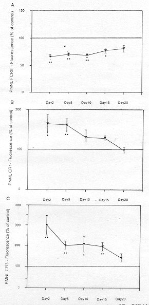
PML- expression of Fey RIII
The patient PM1- expression of Fey RIII was decreased to
65.7% of control values at admission and remained low for the first 20 days (Fig. M,
Table II). PM1- expression of CRs
The patient PM1- expression of CRI increased to 162.9% of control values at 2 days, and
then gradually decreased to control levels at 20 days (Fig. ]B, Table II).
|
Fig. 1 -
Time course of neutrophil (PML) expression of Fcy RIII (A), CRI (B) and CR3 (C) in burn
patients as determined by indirect immunofluoresence and FM The results are expressed as
the percentage of the fluorescence obtained with PMLs from normal subjects in parallel
measurements. The results are given as the mean ± SEM and significance of difference
between the patients and controls is indicated as *p < 0.05, ** p < 0.001. |
|
| |
Fey R111 |
CRI |
CR3 |
| Controls |
2.9 ± 0.3 |
1.9 ± 0.12 |
6.6 ± 1.6 |
Patients:
(days post-burn) |
|
|
|
2 |
1.86 ± 0.13** |
3.1 ± 0.43* |
19.2 ± 3.3** |
5 |
1.98 ± 0.1** |
3.0 ± 0.3** |
12.7 ± 1.4** |
10 |
1.94 ± 0.11 * |
2.4 ± 0.55 |
12.9 ± 2.6* |
15 |
2.2 ± 0.13* |
2.5 ± 0.21 |
12.1 ± 1.4** |
20 |
2.3 ± 0.2 |
1.86 ± 0.16 |
8.3 ± 0.98 |
|
| Table II - PIVIL expression of
Fey RIll, CRI and CR3 as determined by immunofluoresence and FCM in 23 burn patients and
10 controls. The results are given as the mean ± SEM (%). The significance of difference
between patients and controls is indicated as *p < 0.05, ** p < 0.001 |
|
The expression of
PM1- CR3 increased by 199% on day 2 and remained high during the first 20 days (Fig. X,
Table II). Both control and patient PN1L CRI-dependent and CR3-dependent fluorescence
were shown to be monophasic throughout the investigation period.
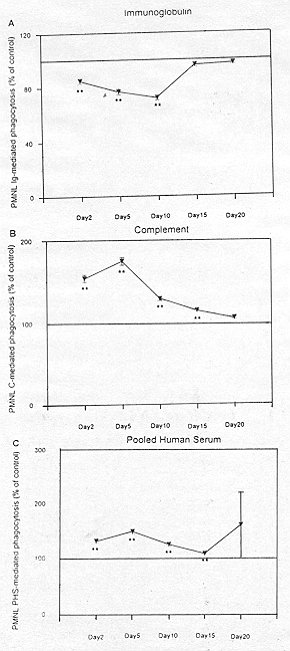
Phagocytosis
The PML Ig-mediated phagocytosis of C. albicans
decreased to 84.7% of control values on day 2. The lowest Ig-mediated phagocytosis was
observed on day 10, with a reduction of 28.1% compared with controls (Fig. 2A, Table
III).
The patient PML complement-mediated phagocytosis of C. albicans was increased by about
54% of control level and remained high for the first 20 days (Fig. 213, Table III).
The patient PML phagocytosis of C. albicans opsonized with PHS (i.e. in the presence of
both IgG and complement) increased by about 3 1 % of control values and remained higher
than that of control for 20 days (Fig. 2C, Table III).
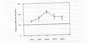
Plasma levels of ICAM-1
Plasma ICAM-1 increased by 31% of control values on day 2 and remained high for the first
20 days (maximum level on day 10 (Fig. 3, Table IV).
Discussion
Neutrophil activation by chemoattractants, enzymes and
bacteriologically-derived peptides release Pl-linked FeRIll into plasma and in tissues
with active inflammation.'PI-Iinked FcRIll mediates exocytosis of neutrophil granule
proteins but does not mediate the initiation of the respiratory burst.` The present study
demonstrates that expression of PML Fcy RIII following burn injury decreased on day 2 and
remained low for the first 20 days. This result is thus indicative of a systemic
activation of these cells, which induce shedding of this receptor, a finding in agreement
with Vindenes and Bjerknes.
|
Fig. 2 -
Time course of neutrophil (PIVIL) immunoglobulin-mediated phagocytosis (A),
complement-mediated phagocytosis (B), and phagocytosis mediated by pooled human serum
(PHS) (C) in burn patients as determined by phagoeyfic index. The results are expressed as
the percentage of the number of Candida albicans ingested by 100 PMLs divided by
100. The control data were obtained from parallel measurements. The results are given as
the mean ± SEM, and the significance of difference between the patients and controls is
indicated as *p < 0.05, **p < 0.01. |
|
| |
lg-mediated
phagocytosis |
Complement-mediated
phagocytosis |
PHS-mediated
phagocytosis |
| Controls |
95.3 ± 3.9 |
94.5
± 2.1 |
118.8 ± 2.3 |
Patients
(days post-burn) |
|
|
|
2 |
79.6 ± 2.0** |
147.4
± 3.6** |
156,8 ± 4.5** |
5 |
71.5 ± 2.1** |
167.4
± 3.3** |
178.7 ± 4.9** |
10 |
66.9 ± 1.1** |
123.0
± 1.9** |
150.7 ± 3.9** |
15 |
91.3 ± 1.8 |
109,3
± 1.1** |
129.3 +-1.2** |
20 |
92.6 ± 1.1 |
100,2
± 1.8 |
194.5 +-76.5 |
|
| Table III - PM1-
opsonophagocytosis mediated by immunoglobulin, complement and pooled human serum
determined by phagocytic index in 23 burn patients and 10 controls. The results are given
as the mean ± SEM (%), and the significance of difference between patients and controls
is indicated as *p < 0.05, **p < 0.01 |
|
A marked and sustained increase in the
expression of the complement receptors CRI and CR3 was documented in the present study,
The increase in fluorescence that depends on complement receptors was always observed as a
monophasic peak, indicating that all the cells had been activated by a stimulus that was
systemic. Moore et al." and Vindenes and BjerkneS26 reported similar results. The
increased expression of CR3 is an important part of neutrophil priming and activation as
this molecule is critically involved in adherence of PML to the endothelium. This is a
necessary initial step in the emigration of PMLs from the circulation into the tissues. In
addition, both CRI and CR3 are necessary for optimal phagoeytosis of complement-coated
bacteria and immune complexes.
| |
ICAM- 1 |
| Controls |
223.9 ± 21.7 |
Patients
(days post-burn) |
|
2 |
298.3 ± 23.2 |
5 |
367.5 ± 43. 1 |
10 |
507.5 ± 8.9** |
15 |
392.5 ± 56.6 |
20 |
382-2 ± 284** |
|
| Table 1V - Plasma levels of
ICAM-1 as measured by ELISA in 23 burn patients and 10 controls. The results are expressed
as the mean ± SEM (ng/ml), The significance of difference between patients and controls
is indicated as *p < 0.05, **p < 0.01 |
|
Previous studies
have shown that a variety of mediators such as the complement split product C5a '27
TNF-(1,28 IL-8,' granulocyte macrophage colony-stimulating factors' and endotoxin,"
which might be locally produced at sites of infection, are all capable of increasing
complement receptor expression.
|
Fig. 3 -
Time course of ICAM-1 plasma levels in burn patients determined by ELISA. The results are
expressed as the mean ± SEM (ng/ml). The control data are obtained from parallel
measurements. The significance of difference between the patients and the controls is
indicated as *p < 0.05, **p < 0.01. |
|
There are intracellular pools for both CRI
and CR3, but the intracellular locations for these pools are distinct. The pool for C3
co-sediments with specific granules, while the pool for CRI does not."The increased
receptor expression occurs within minutes and represents a translocation of presynthesized
receptors from intracellular pools to the surface rather than a new synthesis.
Consequently, the increased expression of PML CRs following thermal injury strongly
suggests PML degranulation. This is consistent with earlier reports by Alexander" and
Davis et al.
This study revealed that plasma ICAM-1 levels were elevated during the first 20 days
post-burn, with a maximum level on day 10. This finding could be related to the activation
of the inflammatory cytokines such as IL-16, TNF-P and INF-a, which induce or enhance the
expression of both membrane and soluble ICAM-l."," Jaeschke et al."
reported an increased plasma ICAM-1 level in endotoxin-challenged mice. In acute and
chronic inflammatory processes, fibrin deposition and leukocyte accumulation are classic
histopathological hallmarks. Fibrin deposition on vascular endothelial cells (EQ can
result in the upregulation of EC ICAM-1, which is an important ligand/receptor for
CDllb/CDl8 expressed on neutrophils. Fibrin stimulation of EC increased their adhesiveness
for PMLs.` Circulating levels of soluble endothelial cell adhesion molecules may reflect
the magnitude of expression of their membrane-bound counterparts." Mulligan et
al." have emphasized the role of ICAM-1 in the events that lead to
neutrophil-mediated vascular injury of dermis and lung after thermal trauma to the
skin. These would cause cessation of neutrophil movement along the endothelial cells and
transmigration.
Systemic activation of PMLs may have harmful effects on the host. Increased PM1- CR3
expression as well as ICAM-1 plasma level may cause increased neutrophil adhesiveness and
the formation of leukoeyte microemboIi that concentrate in the first capillary bed
encountered, the lung There is also depression of neutrophil chemotaxis.` Activated
PMLs are known to degranulate and release lysosomal enzymes as Well as oxygen radicals
toxic to surrounding tissues. Injury~related PNNI activation can sustain and be
further strengthened by subsequent superimposition of infectious foci (e.g. burn wound
sepsis" or by transintestinal transport of LPS or micro-organisms .
This recruitment and activation of PMLs in the course of thermal injury may exceed
physiological needs and induce PN1L tissue infiltration and destruction. We therefore
recommend that the apparent source of neutrophil-activating substances, the burn wound,
should be excised as early as clinically feasible in order to free the patient of the
burden of systemically activated neutrophils` and early initiation of enteral feeding. The
use of anti-proinflammatory cytokines and/or anti-ICAM-1 needs further investigation.
RESUME. Les leucocytes
polymorphnucléaires (LPMs) jouent un rôle fondamental dans la défense de l'hôte, et le
dysfonctionnement phagocytaire a été associé à la susceptibilité augmentée à
l'infection dans les patients brûlés. La molécule-1 d'adhésion intercellulaire joue un
rôle dans l'accumulation leucocytaire et l'extravasation. L'analyse cytométrique du flux
a été utilisée pour étudier l'expression par les LPMs du récepteur Fe 111 IgG (Fcy
RIII) comme aussi des récepteurs du complément CRI (récepteur pour C3b) et CR3
(récepteur pour C3bi) dans 23 grands brûlés. Cette étude a indiqué une augmentation
significative, avec une certaine variation, de la fluorescence, de la phagocytose obtenue
avec le complément de Candida albicans, et du plasma ICAM, après le deuxième
jour et pour les vingt jours suivants. La fluorescence dépendante de la FCy RIII et la
phagocytose obtenue avec l'Ig démontraient, au contraire, une augmentation significative
par rapport aux valeurs témoins. Ces résultats indiquent une variation significative
dans l'expression du récepteur de l'opsonine des LPMs et dans la phagocytose, ce qui
confirme l'activation systémique des LPMs après les grandes brûlures. En outre,
l'élévation de lICAM-1 plasmatique peut augmenter l'effet nuisible de l'activation des
neutrophiles.
BIBLIOGRAPHY
- Polk H.C.: Consensus summary on infection. J. Trauma, 19:
894, 1979.
- Ninnemann J.L.: Immunologic defenses against infection:
Alterations following thermal injury. J. burn Care Rehabil., 3: 355, 1982.
- Klebanoff S.J., Clark R.A.: "The Neutrophil: Function
and Clinical Disorders". Amsterdam, North-Holland Pub]., 409-88, 1978.
- Unkeless J.C., Wright S.D.: Phagocytic cells: Fey and
complement receptors. In: "Inflammation: Basic Principles and Clinical
Correlates". Gallin J.1., Goldstein I.M., Snyderman R. (eds), New York, Raven Press,
343-62, 1988.
- Neuman E., Huleatt JW., Jack R.M.: Gramdocyte-macrophage
colony-stimulating factor increased synthesis and expression of CRI and CR3 by human
peripheral blood neutrophils. J. Immunol., 145:3325,1990.
- Detmers P.A., Fowell D.E., Walz A. et al.: Differential
effects of neutrophil-activating peptide-I/IL-8 and its homologues on leukocyte adhesion
and phagocytosis. J. Immunol., 147: 4211, 16.1991.
- Felzmann T., Gadd S., Majdic 0. et al.: Analysis of
function-associated receptor molecules on peripheral blood and synovial granulocytes from
patients with rheumatoid and reactive arthritis. J. Clin. Immunol., 11: 205, 1991.
- Ravetch J., Perussia B.: Alternative membrane forms of
FcR111 (CD16) on human NK cells and neutrophils. J. Exp. Med., 170: 481, 1989.
- Fearon D.T., Wong W.W.: Complement ligand-receptor
interactions that mediate biological responses. Ann. Rev. Inummol., 1: 243, 1983.
- Wright S.D., Rao P.E., Van Vorrhis W.C. et al.:
Identification of the C3bi receptor of human monocytes and macrophages using monoclonal
antibodies. Proc. Nail. Acad. Sci. USA., 80: 5699, 1983.
- Berger M., O'Shea J., Cross A.S. et al.: Human neutrophils
increase expression of C3bi as well as C3b receptors upon activation. J. Clin. Invest.,
74: 1566, 1984.
- Springer T.A.: Adhesion receptors of the immune system.
Nature, 346: 425, 1990.
- Newman W, Beall L.D., Carson CW. et al.: Soluble E-selectin
is found in the supernatant of activated endothelial cells and is elevated in the serum of
patients with septic shock. J. Immunol., 150: 644, 1993.
- Lambe J.R., Skinner M.P., Berndt M.C. et al.: Prevention of
activated neutrophil adhesion to endothelium by soluble adhesion protein GMP 140. Science,
249: 414, 1990.
- Dustin M.L. Rothlein R., Bhan A.K. et al.: Induction by
IL-I and interferon-y: tissue distribution, biochemistry and function of a natural
adherence molecule (ICAM-1). J. Immunol., 137: 245, 1986. Ballantyne C.M., Mainolfi E.A.,
Young J.B. et al.: Prognostic value of increased levels of circulating ICAM-1 after heart
transplantation. Clinical Research, 39: 286a, 1991.
- Seth R., Raymond F.D., Makgoba, M.W.: Circulating ICAM-1
isoform diagnostic prospects for inflammatory and immune disorders. Lancet, 338: 83, 1991.
- Nelson R.D., Hasslen S.R., Ahrenholz D.H. et al.:
Mechanisms of loss of human neutrophil chemotaxis following thermal injury. J. burn Care
Rehabil., 8: 496, 1987.
- Grogan J.B.: Altered neutrophil phagocytic function in burn
patients. J. Trauma, 16: 734, 1976.
- Heck E.L., Edgar M.A., Masters B.S. et al.: The role of
NADHNADPH oxidase activity in the leukocyte function of burned patients. J. Trauma, 19:
49, 1979.
- Alexander J.W., Wixon D.: Neutrophil dysfunction and sepsis
in burn injury. Surg. Gynecol. Obstet., 130: 431, 1970.
- Duque R.E., Phan S.H., Hudson J.L. et al.: Functional
defects in hagocytic cells following thermal injury: Application of flow cytometric
analysis. Am. J. Pathol., 118: 16, 1985.
- Ballart I.J., Estovez M.E., Diez R.A., Sen L.: Comparison
of Candida-killing activity measured by chemiluminescence and cytomorphological methods in
human phagocytes. J. Immunol. Meth.,97: 263, 1986.
- Huizinga T.W.J., de Hass M., Kleijer M. et al.: Soluble Fcy
receptor 111 in human plasma originates from release by neutrophils.Clin. Invest., 86:
416, 1990a.
- Huizinga T.W.J., Dolman N.J., Van der Linden M. et al.:
PI-linked FcR I I I mediates exocytosis of neutrophil granule proteins, but does not
mediate initiation of the respiratory burst. J.Immunol.,144: 1432, 1990b.
- Vindenes H., Bjerknes R.: Activation of polymorphnuclear
neutrophilic granulocytes following burn injury: Alteration of Fc-receptor and
complement-receptor expression and of opsonopha gocytosis. J. Trauma, 36: 161, 1994.
- Moore F.D., Davis C., Rodrick M. et al.: Neutrophil
activation in thermal injury as assessed by increased expression of complement receptors.
N. Eng. J. Med., 314: 948, 1986.
- Berger M., Wetzler E.M., Wallis R.S.: Tumor necrosis factor
is the major monocyte product that increases complement receptor expression on mature
human neutrophils. Blood, 71: 151,1988.
- Davis C.F., Moore F.D., jr, Rodrick M.L. et al.: Neutrophil
activation after burn injury: contributions of the classic complement pathway and of
endotoxin. Surgery, 102: 477, 1987.
- O'Shea J.J., Brown E.J., Seligmann B.E. et al.: Evidence
for distinct intracellular pools of receptors for C3b and C3bi in human neutrophils. J.
Immunol., 134: 2580, 1985.
- Alexander JW.: Serum and leukocyte lysosomal enzymes:
Derangements following severe thermal injury. Arch. Surg., 95: 482,1967.
- Davis J.M., Dineen P., Gallin J.I.: Neutrophil
degranulation and abnormal chemotaxis after thermal injury. J. Immunol., 124: 1467, 1980.
- Pober J.H., Lapierre L.A., Stolpen A.H. et al.: Activation
of cultured human endothelial cells by recombinant lYmphotoxin: comparison with tumor
necrosis factor and interleukin-I species. J. Immunol., 138: 3319, 1987.
- Jaeschke H.N.A., Essam M.A., Fisher S.L. et al.: Release of
soluble intercellular adhesion molecule- 1 into bile and serum in murme endotoxin shock.
Hepatology, 23: 530, 1996.
- Qi J., Kreutzer D.L., Piela-Smith T.H.: Fibrin induction of
ICAMI expression in human vascular endothelial cells. J. Immunol., 158: 1880, 1997.
- Henninger D.D., Panes J., Eppihimer M. et al.: Cytokine
induced VCAM-1 and ICAM-1 expression in different organs of the mouse. J. Immunol. 158:
1825, 1997.
- Mulligan M.S., Till G.O., Smith CW. et al.: Role of
leukocyte adhesion molecules in lung and dermal vascular injury after thermal trauma of
skin. Am. J. Path., 144: 1008, 1994.
- Ward P.A., Till G.O.: Pathophysiologic events related to
thermal injury of skin. J. Trauma, 30 (Suppl.): 575, 1990.
- Winchurch R.A., Thupari J.N., Munster A.M.: Endotoxemia in
burn patients: Levels of circulating endotoxins are related to burn size. Surgery, 102:
808, 1987.
- Ziegler T.R., Smith R.J., O'Dwyer S.T. et a].: Increased
intestinal permeability associated with infection in burn patients. Arch. Surg., 123:
1313, 1988.
- Meyer J., Yurt R.W., Duhaney R. et al.: Differential
neutrophil activation before and after endotoxin infusion in enterally versus parenterally
fed volunteers. Surg. Gynecol. Obstet. 167:501, 1988.
Moore F.A., Moore E.E., Jones T.N. et
al.: TEN versus TPN following major abdominal trauma reduced septic morbidity. J. Trauma,
29: 916, 1989.
This paper was
received on 5 November 1997
Address correspondence to:
Dr Samy A. Shehab El-Din
Plastic, Reconstructive and burn Unit
Mansoura University Hospitals
Mansoura, Egypt
fax: 2050 348203 |
|