Annals of Burns and Fire Disasters - vol. XI
- n. 3 - September 1998
STRESS ANALGESIA
ASSOCIATED WITH EXPERIMENTAL BURN SHOCK
Sinitsin L. N.
Research Institute of
Traumatoiogy and Orthopaedics, Ministry of Health, Nizhniy Novgorod, Russia
SUMMARY. This
paper considers excitation transmission changes in the afferent and visceral efferent
cerebral systems, the threshold of pain sensitivity, the endogenous opiate synthesis
level, and the character of the antishock agent activity due to burn shock.
Introduction
Experiments with curarized
and moderately anaesthetized adult cats with burn shock have demonstrated a pronounced
depression of excitation transmission in the associative and nonspecific afferent systems
during somatic, sound and light stimulation. The effects controlling the activity of the
cardioavascular system are facilitated in the efferent systems. In white rats, burn shock
leads to an increase in the somatic and visceral pain threshold during the first five
days. Rausedyl and naloxone reduces stress analgesia caused by the burn shock in white
rats. The burn shock enhances opiate-like activity (beta-endorphin, beta-lipotropin) of
the white rat forebrain, as shown by radioimmunoassay. The data suggest that stress
analgesia associated with experimental burn shock is likely to be accounted for by the
increased production of endogenous opiates.
CNS disturbances due to the burn shock are well known.' However, the peculiarities of
excitation transmission changes in cerebral structures due to shock of various aetiology
have not been well investigated. This matter has recently aroused great interest owing to
the development of the endogenous opiate theory, according to which the vital activity
products of the brain regulate its excitability and influence impulse transmission,
causing stress analgesia in extreme conditions.
The present study investigates excitation transmission changes in the afferent and
visceral efferent cerebral systems, the threshold of pain sensitivity, the endogenous
opiate synthesis level, and the character of the antishock agents activity due to burn
shock.
Methods
The experiments were
carried out on 50 adult cats and 300 intact rats. The cats were anaesthetized with
chloralose (10-30 mg/kg) and immobilized with succinylcholine, with artificial ventilation
control and body temperature within the norm.
The influence of the damaging factors on the excitation transmission in the afferent
systems was investigated with the method of the registration of evoked potentials in
specific associative and non-specific structures of the cerebral hemisphere cortex, the
diencephalon and the mesencephalon, due to somatic, sound and light stimulation.
The evoked potentials were recorded by means of a 6channel "Alvar" amplifier and
a 6-channel cathode-ray oscillograph, using the waiting/scanning system of multiple
superposition on the film. The sciatic nerve was stimulated with single or double I msec
square wave impulses at I imp/sec frequency. Sound and light impulses were received with
corresponding transformations of the electric impulses.
The influence of burn trauma on the excitement transmission in the efferent visceral
systems was investigated under the same conditions with the method of local electrical
stimulation of the sensorimotor zone of the cerebral hemisphere cortex, diencephalon and
midbrain structures and recording of neurogenous arrhythmia, hypertension reactions, and
changes in the general level of arterial blood pressure. ECG in the second standard
conduction and arterial blood pressure in the femoral artery were recorded, by means of
"Cardiovar" and "Bariovar" (manufactured by Alvar). The brain was
stimulated with 1 msec square wave electrical pulses of 5-10 V amplitude with 200 imp/sec
frequency at 5 sec intervals.
A third-degree burn shock (a-b) was inflicted in 20-30% of the total body by contact burn
using a metal plate heated to 100°C.
Results
The results of the
experiments showed that some relief of the excitement transmission in the afferent system
appeared during the first hours after the infliction of the burn trauma in the animals.
The amplitude of the primary responses was enlarged by somatic, sound and light
stimulation in the fields of specific projection in the cerebral cortex. In the
associative and non-specific fields, the amplitude and the length of responses increased
in conditions of sound and light stimulation. On the level of the diencephalon and
midbrain, in the associative and nonspecific thalamus nuclei, the hypothalamus and the
reticular formation structures, the increase in amplitude of the evoked potentials was
observed to be less expressed by somatic stimulation and more markedly increased by sound
and light stimulation. Starting from the second hour, the relief in excitation
transmission modified to suppression, which increased constantly and reached its maximum
at the end of the first 24 h. This was manifested in decreased amplitude and increased
latent period and evoked potential length. The responses in the associative and
non-specific areas of the cerebral cortex were more suppressed and the primary responses
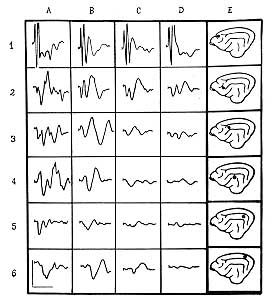
in the fields of specific projection were less suppressed (Fig. 1). In the experiments
with double pulses the responses to the second impulse were mostly suppressed, which
testified to decreased liability of the brain afferent systems.
|
Fig.
I - Influence of burn shock on evoked potentials of cerebral cortex by sciatic nerve
stimulation.
A = initial background.
B = 10 min post-burn.
C = 10 h post-burn.
D = 18 h post-burn.
E = localization of recording electrodes:
1 - specific projection area;
2, 3, 5 - associative projection zone;
4, 6 - non-specific projection zone)
Vertical lines = 100 mkV amplitude;
horizontal lines = time 100 msec. |
|
Analogous changes in
excitation transmission were observed in the associative and nonspecific structures of the
diencephalon and midbrain. In response to sciatic nerve sound and light stimuli, the
amplitude of the evoked potentials decreased, and their latent period and length
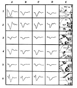
increased. Acute depression of the potentials was observed at the end of the first 24 It (Fig.
2).
|
Fig.
2 - Influence of the burn shock on the evoked potentials of the diencephalon and
midbrain structures.
A = initial background.
B = 10 min post-burn.
C = 10 h post-burn.
D = 18 h post-burn.
E = localization of the recording electrodes:
1 - associative projection area of the thalamus: nucleus medialis dorsalis (MD);
2 - non-specific projection nucleus commissura posterior (NCP);
3 - formatio reticularis mesencephalica (Ret Mes);
4 - colliculus superior (CS);
5 - Ret Mes;
6 - Griseurn Centrale (GC).
Vertical lines -- 100 mkV amplitude;
horizontal lines = time 100 msec. |
|
In the efferent systems
the burn trauma caused an acute increase in the decreased effects controlling the activity
of the cardiovascular system. The general level of arterial blood pressure increased on
average by 18 % during the first hours. Hypertension was maintained in most of the
experiments during the first 24 h, which was followed by and a decrease in the general
level of arterial blood pressure to initial and lower values.
Hypertensive reactions and the stimulation of the sensorimotor areas of the cerebral
cortex nonspecific stiruclures of the diencephalon and midbrain increased on average after
burn infliction by 81% as compared with the initial values and remained at that level
during the first 24 h, after which their intensity gradually decreased.
The continuity of neurogenic arrhythmias resulting from the local stimulation of the
cerebral structures in question increased in 3.7 times during the first 2 h postburn. This
effect gradually decreased but persisted for the first 24 h.
Discussion
A comparative analysis of
the data allowed us to hypothesize that the burn shock caused changes in excitation
transmission in the afferent and efferent brain systems analogous to those observed
following by the action of narcotic analgesics of the morphine group.` These assumptions
formed the basis for further experiments in which the influence of the burn shock on
somatic and visceral pain sensibility was studied.
The experiments were carried out on white rats weighing 150-200 g. The pain thresholds
were determined with the previously developed method of somatovisceral analgesiometry.`
After immobilization the animals received stimulating bipolar electrodes in the upper
third of the tail and abdomen. Stimulation was carried out with 1 msec square wave pulses
in packets (10 impulses per packet) at 5 see intervals. The pain threshold value was
determined by the power value of the stimulating pulse curve causing motor reaction,
squeaking during stimulation, and squeaking after stimulation. The stimulating current
power was measured with a Cl-19 cathode oscillographer by the tension drop on the resistor
switched on consecutively with the object.
The results showed that 1 h post-burn there was an increase in the somatic and visceral
pain sensibility threshold. This gradually increased and reached its maximum level by the
end of the first 24 h, 5-10 times higher than the initial level. The somatic pain
threshold increased in this period more than the visceral threshold. During the following
five days the pain thresholds gradually decreased to initial values.
These data made it possible to assume that the burn trauma caused stress analgesia in
animals which, according to reports, 2-6 was the result of the hyperproduction of
endogenous opiates in the brain structures. To confirm this assumption, we determined the
total opiate-like activity (beta-endorphin, beta-lipotropin) of the anterior cerebral
areas of white rats due to the burn trauma. The investigation was carried out using
N-Nuclear Corporation radio-immune diagnostic sets (USA). Radioactivity was determined by
automatic LKB gamma counter (Sweden).
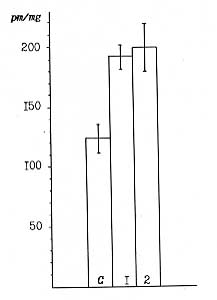
It was established that in the control group of the intact rats the total opiate-like
activity was 121.64 ± 11.9 pm/mg, while in white rats killed 24 h after burn infliction
the opiate-like activity was 199.5 ± 21.6% pm/mg. The results of the experiments
testified to the fact that the burn shock caused opiate-like activity enchancement as
compared with the control values at 75.5 pm/mg in 2 h and 77.55 pm/ing in 24 h post-burn
trauma (p < 0.05) (Fig. 3).
|
Fig.
3 - The effect of burn shock on the total opiate-like activity of the cerebral frontal
areas in white rats.
C = Control group.
1 - 2 h post-burn;
2-24 h post-burn. |
|
On the basis of these
experimental data, we can state that in conditions of experimental hum shock there was
evident stress analgesia resulting from increased production of endogenous opiates.
For further analysis of the stress analgesia mechanism due to burn shock, we used this
model to study the effect of the antagonist agents of the endogenous opiates - naloxone,
rausedyl` and their synergists (narcotic analgesics and enkephalin derivatives) on pain
threshold changes in burned animals.
The results showed that rausedyl in doses of 20 mg/kg reduced the increase of the pain
thresholds of somatic and visceral pain sensibility due to burn shock. Under the same
conditions (5 mg/kg) naloxone also reduced the post-burn increase of the pain threshold.
Action was manifested 10 min after administration and lasted for 1 h. On the second day
the pain thresholds were sharply decreased compared with animals not receiving naloxone (Fig.
4).
Analgesics were administered to white rats before and 10, 30, 60 and 120 min after
infliction of burn trauma, as also after 24-48 h. Morphine (10 mg/kg), phentanyl (10
mk/kg) and lexir (1 mg/kg) administered 2-3 h after the burn trauma were found to cause a
sharp increase of the pain threshold in comparison with both burned and intact animals. In
the latter case, the somatic pain threshold increased respectively 10, 3 and 4 times and
the visceral pain threshold 1.5, 4 and 5 times. The administration of 200 mg/kg gamma-OH
produced a similar effect.
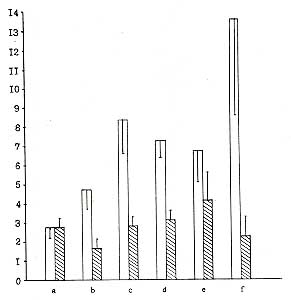 |
Fig.
4 - Influence of naloxone on visceral stress analgesia due to experimental burn
shock in white rats.
Ordinate axis: pain threshold values (in cond. units). Initial pain threshold on basis of
vocalization reaction after stimulation is considered as follows:
a - within 2 min of burn infliction;
b - within 10 min;
c - within 30 min;
d - within 60 min;
e - within 2 h;
f - within 24 h of naxolone administration at dose of 5 mg/kg.
Light columns - control animals, receiving sodium chloride solution;
Shaded columns - animal receiving naloxone (10 rats per group).
Vertical lines indicate confidence limits. |
|
The same doses of the
drugs applied before or 10, 30 and 60 min after burn application were also found to
prevent further increase of the pain threshold after termination of action. After 24 h the
pain thresholds in animals that had received analgesics before or for 1 after the burn
trauma were 3-5 times lower in
comparison with the burned animals in the control group.
The experimental results testify that stress analgesia develops, followed by burn shock, as a result of hyperproduction of
endogenous opiates. These potentiate the analgesic effect of the opiates if the opiates
are applied 2 h after the burn trauma or later. Also, the administration of narcotic
analgesics, either preliminarily or for 1 h, can prevent initiation of the hyperproduction
of endogenous opiates, which is a factor of their antishock effect. Separately we show that ley-enkephaline administration to the brain
ventricles of white rats (1 ing in 5 ml) resulted in the acute increase of the somatic and
visceral sensibility pain thresholds. This demonstrated the role of endogenous opiates in
the development of central analgesic action.
Conclusions
The results of the
investigations testify that excitation transmission in the afferent and efferent cerebral
systems significantly changed in conditions of extreme shock. This led to a modification
in the action of neurotropic agents applied in anaesthesiology in order to prevent and
treat burn shock. The character of the neurotropic agent action varied in relation to the
phase and the degree of the shock. These factors should be taken into consideration in
clinical practice in order to provide more rational and effective correction of
disturbances of the central nervous system in shock conditions.
RESUME.
L'Auteur considčre les modifications de la transmission de l'excitation des systčmes
cérébraux afférent et efférent, le seuil de la sensibilité ŕ la douleur, le niveau
de la synthčse de l'opiacé endogčne, et le caractčre de l'activité des agents
antichoc due au choc de la brűlure.
BIBLIOGRAPHY
- Saacov B.A., Bardakhchyan
E.A.: Vital problems of the burn shock pathogenesis. 1979.
- Golanov EN., Parin S.B., Jasnetsov
V.V..: The effect of nalorphine and naloxone on the electric shock in rabbits. Bull,
Exper. Biol. Med., 6: 60-1, 1992.
- Holaday J.W., Belenky G.L., Faden
A.I. et al.: Possible function of beta-endorphin. Neuropsychopharmacology, Vienna: 503-14,
1978.
- Kelly D.D., Bodnar R.I.: Acupunct.
Elec. They. Res., 4: 159, 1979. Ho W.K.K., Wong H.K., Wen H.L.: The influence of
electroacupuncture on naloxone-induced morphine withdrawal.
- The effect of cyclic-AMP.
Neuropharmacology, 11-18: 865-9, 1982.
- Fadeh J., Holaday J.: J.
Pharmacol. Exp. Ther., 212: 441, 1980.
- Sinitsin L.N.: On the effect of
neurotropic agents on the afferent and efferent visceral systems. Author's abstract of
M.D. dissertation, 1974.
- Sinitsin L.N., Ivanov A.M.: The
effect of the burn shock on the excitation transmission in the afferent and efferent
celebral systems. The problems of the burn pathology. Collection of scientific papers,
GRITO, Gorky: 158-61, 1980.
- Sinitsin L.N., Gelashvili S.S.,
Maksimov G.A.:. Neurophysiological aspects of shock condition. Neurodynamics of the brain.
Trauma. Collection of scientific papers, GRITO, Gorky, 197-209, 1984.
- Zakusov V.V.: Pharmacology of
central synapses. 116, 1973.
- Sinitsin L.N.:. Rationalization
proposal. 645, GRITO, 1980.
This paper was received
on 10 January 1997.
Address correspondence to:
Dr L.N Sinitsin
Research Institute of Traumatology and Orthopaedics
Ministry of Health
Nizlymy Novgorod, Russia. |
|