Annals of Burns and Fire Disasters - vol. X1 - n. 3 - September 1998 INTERDENTAL WIRE FIXATION OF ENDOTRACHEAL TUBE FOR SURGERY OF SEVERE FACIAL BURNS Botts J., Srivastava K.A., Matsuda T, Hanumadass M.L. Sumner L. Koch Burn Center, Cook County Hospital and University of Illinois College of Medicine, Chicago, Illinois, USA SUMMARY. Complex facial burns requiring complete exposure of the face for initial skin grafting and secondary reconstructive surgery prompted our improvising a wire fixation method of securing the oral endotracheal tube. Utilizing this method, we have experienced no accidental extubations or trauma to the patient's dentition. A small amount of gingival bleeding is possible both during and after insertion and removal of the wire. Alternative methods must be utilized for both edentulous patients and those with prosthetic dental devices located in the anterior portion of the mouth. We have found this method to be superior to the external cranial fixation device of Hansen, the lip fixation method, and various taping techniques when diffuse severe facial burns require reconstructive surgery. Not only does the wire fixation method appear applicable to a wider range of facial surgery such as diffuse burns or severe trauma but the fact that the tube can be safely secured in 5-10 minutes gives an advantage over other previously described methods. Introduction Diffuse burns of the face have long been a challenge to the burn surgeon, both in surgical technique and in exposure for the surgical procedure. Facial surgery of any type usually presents a problem with securing of the endotracheal tube: the securing tape impinges on the operative field.` Many methods and devices have been constructed for fixation of the endotracheal tube for various types of surgical procedures on the face. Two of the previously utilized methods are external cranial frame fixation' and the securing of the endotracheal tube to the lower lip with nonabsorbable sutures. None of these procedures is satisfactory in every case. We have devised a method using dental wire to secure the endotracheal tube for acute and reconstructive facial surgery of patients with severe burn injuries. Material and methods After oral intubation with
a standard endotracheal tube, the tube is connected to a corrugated metal connection
extension (gooseneck adapter) to allow better accessibility and exposure of the entire
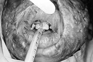
operative field. An eight-to-ten inch segment of number twenty-five dental wire is used,
held by one end with a standard needle holder. The wire is first positioned by an anterior
approach between the interdental spaces of the first and second incisors on one side of
the upper teeth. Next, through a posterior approach, the interdental space of the first
and second incisors on the opposite side of the upper teeth is entered, resulting in the
two ends of the wire exiting anteriorly. It is imperative to include two teeth in the wire
loop, either central incisors or the first and second incisors, on either side of the
upper dentition.
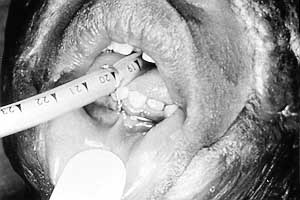
It is necessary to ensure that the tube is unable to be moved anteriorly or posteriorly with moderate traction once secured. The excess wire is then cut and the ends bent upon themselves to avoid lacerations or punctures of the oral mucosa. If the upper teeth are loose or missing, the endotracheal tube can be positioned in a similar manner to the lower teeth. Since the normal mobility of the mandible allows for exaggerated movement of the secured tube in both an anterior and a posterior direction despite proper wire fixation, this method should be used secondarily (Fig. 4).
Discussion The method of securing an
endotracheal tube with dental wire to the upper incisors is a dated, simple, yet not
wellknown procedure which can be performed in approximately five to ten minutes in the
operating room. With the wire fixation method, the wire can be easily removed prior to
reversal of anaesthesia. Removal may be accomplished utilizing either plain wire-cutters
or unravelling the ends simultaneously with two needle holders. Following wire removal and
application of facial dressings, extubation can proceed as planned.
RESUME. Les brűlures complexes faciales qui nécessitent l'exposition complčte du visage pour la greffe initiale de la peau et pour la chirurgie reconstructive secondaire ont stimulé l'intéręt des Auteurs ŕ créer une méthode oů la fixation du tube oral endotrachéal est obtenue moyennant l'emploi d'un fil métallique. Avec cette méthode ils n'ont pas rencontré aucune extubation accidentelle ou traumatisme pour la dentition du patient. Il est possible d'avoir une hémorragie gingivale modérée pendant et aprčs l'introduction et l'enlčvement du fil. Il faut suivre des méthodes alternatives pour les patients édentés et ceux qui ont des prothčses dentaires insérées dans la portion antérieure de la bouche. Les Auteurs ont trouvé que la méthode décrite est supérieure ŕ l'appareil de fixation crânienne externe de Hansen, ŕ la méthode de la fixation ŕ la lčvre et ŕ plusieurs techniques d'attachement avec du ruban, dans les cas des graves brűlures diffuses qui nécessitent la chirurgie corrective. Non seulement la méthode de la fixation moyennant un fil est utilisable dans une gamme plus vaste de la chirurgie faciale mais la possibilité de fixer le tube sans risque en 5-10 minutes présente des avantages par rapport aux autres méthodes décrites. BIBLIOGRAPHY
|