Annals of Burns and Fire Disasters - vol. XI -
n. 4 - December 1998
LYELL'S SYNDROME MANAGEMENT IN A
BURN UNIT.
COVERAGE WITH CRYOPRESERVED ALLOGRAFT
Garcia Bernal F.J.,
Torrero V., Regalado J., Ferdinandez Samaniego F., Gabilondo F. J.
Department of
Plastic and Reconstructive Surgery and Burn Centre, Cruces Hospital, Bilbao, Spain
SUMMARY. Eight
cases of Lyell's syndrome or toxic epidermal necrolysis are described that have been
managed in the Cruces Hospital Burn Centre in the last five years. The therapeutic approach to the
patients is presented, describing the methodology of the systemic treatment and the
disease's cutaneous manifestations. Along with the general treatment scheme, the topical
care of the cutaneous manifestations is presented. Cryopreserved allografts obtained from
our skin bank were used as a skin coverage tool.
Introduction
Clinically, Lyell's
syndrome and the StevensJohnson syndrome or toxic epidermal necrolysis are a severe form
of exfoliative den-natitis accompanied by systemic changes that share certain clinical and
anatornopathological aspects with the disorders caused by intermediate second-degree
burns.
In most cases a pharmacological aetiology is present. Other factors indicated as
aetiological factors in Lyell's syndrome are micro-organisms, food, and other elements
exerting an antigenic action.
In the therapeutic approach, fluid loss replacement and restoration of the dermoepidermic
barrier are priorities, as well as an infection control policy by means of isolation of
the patient. For these reasons, in our experience, such patients could benefit from
management in a burns unit.
Clinical picture
The cutaneous
manifestations of Lyell's syndrome begin as a pruriginous erythema and mild discomfort in
the face and upper thorax, which extend through the extremities within a few days. The
erythema turns into a bullous, exfoliative lesion that is macroscopically similar to a
superficial second-degree scald burn. When there are no complications, this cutaneous
affection tends towards spontaneous wound healing in less than two weeks, with no
scarring.
In its severest forms, the cutaneous affection covers 100% of the body surface.
Characteristically the palms and soles are affected.
The oral, ophthalmic, urogenital and rectal mucosae are typically affected. Perioral
crusting may impair oral nutritional support. Oropharyngeal involvement causes a
troublesome cough. Haernaternesis and melaeria are occasionally seen secondary to
intra-oral, oesophageal, gastric or intestinal mucosal haemorrhages. The gastrointestinal
tract mucosa may be affected at any level, with manifestations such as rectal bleeding and
proctalgia occasionally being present.
Urogenital mucosa involvement causes dysuria and itching. Ophthalmic mucosa involvement
may produce photophobia, conjunctivitis and synechia, which compromise the integrity of
visual function.
The syndrome is characteristically accompanied by fever and severe pain.
Nikolsky's sign, or skin exfoliation with minimal trauma, although not specific, is often
found when we examine these patients.
Histopathology
The final diagnosis is
always defined by skin biopsy. The histological study reveals an eosinophilic necrosis
with subepidermal blisters with a den-noepidermal exfoliation level. There is a minimal
inflammatory response and immunofluorescence tests are negative.
Practical approach to
treatment
Treatment of the condition
has three primary goals:
- haemodynarnic stability
- pain management
- infection control policy
With regard to this last
goal, it is our opinion that isolation of the patient in a burns unit greatly improves
infection control as well as guaranteeing adequate skin coverage.
The management protocol consists of:
- monitoring of various parameters used in
burn patients: pulse rate, arterial pressure, body temperature, central venous pressure,
percentage of oxygen-haemoglobin saturation, urine output, etc.
- laboratory tests to cheek fluid
requirements
- periodic surface cultures of the regions
affected and blood cultures if there is fever above 38.5 °C (101.3 °F)
- management of the patient on an
air-fluidized bed in order to avoid decubitus ulcers and to obtain an adequate temperature
and humidity state
- intravenous access through a central line
for fluid therapy
- enteral nutrition if oral feeding is
impaired, or parenteral feeding if mucosal damage is present
- intravenous narcotics for pain control,
such as morphine 0. 1 rng/kg/24 h
- oral antiacids for gastric stress ulcer
prophylaxis no empirical antibiotherapy is indicated, either systemic or topic. If any
infectious process is present, a specific therapy is applied by means of cultures and
antibiograms
- we do not indicate steroid therapy, in the
light of numerous articles that have found a relationship between a higher mortality rate
and the initiation of this therapy
- the management of the wound is performed
under sedation and analgesia, with Midazolam and Fentanyl, in adequate doses, in relation
to the patient's body weight. Skin cultures are taken, loose devitalized epidermis and
blisters are removed, and the resulting wound is covered with cryopreserved allografts if
the damaged skin exceeds 15% of the body surface
Wound management
First of all a calculation
is made of the affected surface area, reckoning that 1% equals 150-180 cm2. It
is advisable to calculate the injured area periodically, as the initial erythema may
develop into exfoliative lesions, thus becoming an injured area susceptible to coverage.
The cryopreserved allografts proceed from our skin bank and are unfrozen by immersion in
warm water (Fig. 1).
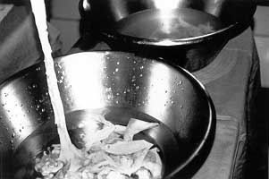 |
Fig.
1 - Allografts from skin bank unfrozen by water- immersion. |
|
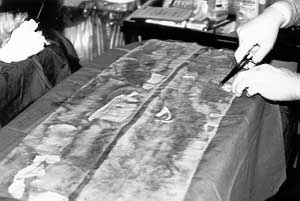
The allografts are applied
on the de-epithelialized areas using an aseptic technique. For fixation of the allografts
we employ a xerofonn and soft gauze dressing (Figs. 2, 3).
 |
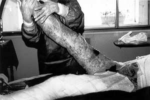 |
Fig. 2 - Allograft placed on sterile gauze before application on
skin. |
Fig. 3 - Allografts applied under aseptic conditions on,
deepithelialized skin, using only xeroform gauze without any fixation system. |
|
The use of an airfluidized bed (Fig. 4) is advised
for the patient's comfort and in order to facilitate wound epithelialization.
The external dressings are changed periodically in relation to clinical evaluations. The
allografts remain attached to their bed until they peel off after about 10-12 days, by
which time the epithelial layers have been restored. If there is any sign of infection,
the allografts must be removed and replaced.
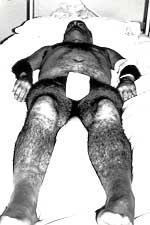 |
Fig. 4
- Air-fluidized bed to facilitate wound epithelialization. |
|
Case reports
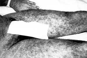
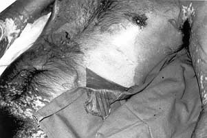
Case 1.
45-yr-old man, twice operated for detachment of the retina. Admitted to the burn unit
after a one-week history of itching, 38 °C fever, and a cutaneous rash involving 90% of
the total body surface area (TBSA), as well as involvement of mucosal surfaces (Figs. 5a,
b). Skin biopsy confirmed Lyell's syndrome. Treatment consisted of fluid infusion, use of
an air-fluidized bed, morphine, and enteral and parenteral support. On the third day after
admission the exfoliative skin was removed and the resulting wound covered with
cryopreserved allografts. Epithelial regeneration was complete, without any sequelae, two weeks after admission.
 |
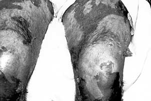 |
Fig. 5a - Case 1. Exfoliative disorder all over body surface. |
Fig. 5b - Case 1. Epidermal exfoliation. |
|
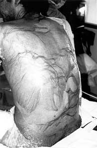
Case 2.
35-yr-old man, HIV-positive, suffering from Pneumocystis carinii, cerebral toxoplasmosis,
oral mycosis, genital herpes, and tuberculosis. After a seizure episode the patient was
treated with phenytoin. 24 h later he began to present itching, fever, a skin rash
involving 90% TBS, and a severe mucosal affection. General measures of patient management
were adopted and on the fifth day after admission the wound was covered with cryopreserved
allografts. Eight days later the skin was completely regenerated (Figs. 6a, b, c, d, e,
f). Skin biopsy diagnosis indicated Lyell's syndrome.
 |
 |
| Fig.
6a - Case 2. Positive Nikol sky's sign. |
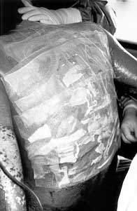
Fig. 6b - Case 2. Allografting. |
|
 |
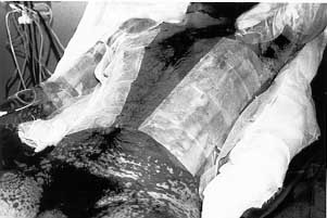 |
| Fig.
6d - Case 2. Exfoliation of skin and positive Nikolsky's sign. |
Fig.
6e - Case 2. Allografting. |
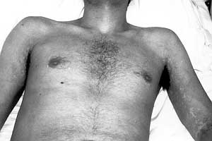 |
Fig.
6f - Case 2. Restitutio ad integrum of skin. |
|
Case 3.
72-yr-old man treated with diuretics, digitalis and allopurinol. Referred 8 days' history
of fever and progressive exfoliative skin disorder affecting 70% TBSA, as well as
Nikolsky's sign and severe mucosal damage. 48 h after admission and administration of
general care measures, allografts were applied for wound coverage. Wound healing was
complete in seven days. Lyell's syndrome confirmed by skin biopsy.
Case 4.
44-yr-old male, HIV-positive in terminal phase, with bronchopulmonary source of sepsis.
When treatment with ceftriaxone was initiated, the patient developed fever, itching, and a
cutaneous exanthema on the face, which in 48 h became a generalized exfoliative skin
disorder with mucosal involvement, the palms and soles also being injured. The patient
died three days after admission, before skin coverage could be performed. Skin biopsy
confirmed the diagnosis.
Case 5.
35-yr-old male, HIV-positive, with herpes zoster, brain tumour, and oesophageal
candidiasis. Treated with phenytoin after a seizure episode. A few hours later generalized
itching developed, together with fever and a blistering skin reaction on the face,
extending to the trunk and extremities. Within 48 h the skin disorder had become general
with a positive Nikolsky's sign. The patient died as a consequence of multi-organic
dysfunction. Skin biopsy indicated Lyell's syndrome.
Case 6.
4-yr-old schoolboy, suffering from fever, itching and a skin rash after trimethoprim -
sulphamethoxazole treatment for a bronchopulmonary process. Within 24 h the rash, which
was first limited to the face, extended to the upper extremities, chest, palms, and soles,
along with mucosal damage. The above-described treatment was established and allografts
were applied. Skin regeneration was complete in 10 days and the patient was moved to the
paediatric area for treatment of the bronchopulmonary process. Skin biopsy confirmed the
diagnosis.
Case 7.
9-yr-old boy treated with carbamazepine for seizures. After a few h, onset of fever and
cutaneous rash in the face. After 48 h the rash became an exfoliative dermatitis in nearly
100% TBSA with severe mucosal damage. After debridement of the blisters the wound was
covered with cryopreserved allografts. Skin completely regenerated 14 days after
admission. Skin biopsy was positive for Lyell's syndrome.
Case 8.
44-yr-old woman admitted to the burns unit suffering from itching, fever, and a facial
rash which progressively extended to the upper extremities and trunk, with severe mucosal
damage. TBSA affected was 45%. The patient had been treated with Ampicillin for an
oropharyngeal affection. Treated as described above, without skin allograft for the wound.
Skin regeneration was complete in 10 days. The patient suffered the complication of
gastrointestinal bleeding and sepsis due to Acinetobacter baumanii. Discharged from
hospital 22 days after admission. Skin biopsy positive for Lyell's syndrome.
Discussion
Toxic epidermal necrolysis
is mainly a severe mucocutaneous exfoliative reaction that requires systemic treatment
along with several topical measures. It is essential in the diagnosis phase to search for
the drug intake antecedent. Cutaneous isolation measures, control of infection, and the
management of cutaneous coverage improve the symptoms and facilitate evolution of the
injuries towards epithelialization. Mucosal hygienic measures are of fundamental
importance to increase the patient's comfort and to prevent the appearance of complications and sequelae. Steroid
therapy is not administered in our burn centre, in line with the experience of other
clinicians. The empirical administration of systemic or topical antibiotics is not
indicated, unless there is a concomitant infectious process, in which case a specific
antibiotic is used, as indicated by the cultures and antibiograms. For wound coverage we
employ a biological skin substitute, cryopreserved allograft, in order to prevent
infection, diminish pain, and prevent fluid losses, thus contributing to homeostasis.
Conclusions
Lyell's syndrome is a
severe disease with a high mortality rate in which systemic treatment, along with
meticulous cutaneous management, is essential. Considering the syndrome's clinical
features, Lyell's syndrome patients would benefit from the management of their pathology
in a burn centre/unit.
In our experience, the cryopreserved allograft is an ideal tool to promote recovery of the
cutaneous barrier because it helps to diminish fluid losses, the intensity of
inflammation, pain, and the risks of infection.
RESUME. Les
Auteurs présentent huit cas du syndrome de Lyell ou la nécrolyse épidermique toxique
qui ont été traités dans le Centre des Brűlures de l'Hôpital Cruces (Bilbao, Espagne)
dans les derniers cinq ans. Ils présentent l'approche thérapeutique employée pour les
patients, avec une description de la méthodologie du traitement systémique et des
manifestations cutanées de la maladie. Ils présentent en outre le plan général du
traitement et les soins topiques pour les manifestations cutanées. Pour la couverture de
la peau ils ont utilisé des allogreffes obtenues dans leur banque de la peau.
BIBLIOGRAPHY
- Napoli B., D'Arpa N., Sferrazza
Papa G., Masellis M.: A case of toxic epidermal necrolysis associated with mycosis
fungoides and complicated with consumption coagulopathy. Ann. Medit. Burns Club, 8: 11-16,
1995.
- Anhalt G., Snelling C.T.F.:
Toxicoepidermal necrolysis. CaseReport. Plast. Reconstr. Surg., 61: 905-10, 1978.
- Ward D.J., Krzeminska E.C., Tanner
N.S.B.: Treatment of toxic epidermal necrolysis and review of six cases. Burns, 16: 77-
t04, 1990. 13.
- Kucan J.V.: Use of Biobrane in the
treatment of toxic epidermal necrolysis. J. Burn Rehabil., 16: 324-8, 1995.
- Prasad J.K., Feller 1,, Thompson
P.D.: Use of amnion for the treatment of Stevens-Johnson syndrome. J. Trauma, 26: 945-6,
1986.
- Pousa F., Valero J., Vdzquez-Barro
A., Trincado S.: Burn unit treatment of three Stevens-Johnson syndrome cases with
cryopreserved allograft. Ann. Medit. Burns Club, 5: 160-3, 1992.
- Napoli B., D'Arpa N., Masellis M.,
D'Amelio L., Genovese M.: Use of cultured homologous keratinocytes in the local treatment
of Lyell's syndrome. Ann. Medit. Burns Club, 9: 163-7, 1996.
- Hemabach D.M., Engrav L. et al.:
Toxic epidermal necrolysis. A step forward in treatment. JAMA, 257: 2171-5, 1987.
- Dending R.H. et al.: Burn unit
management of toxic epidermal necrolysis. Arch. Surg., 113: 758-9, 1978.
- Birchall N. et al.: Toxic
epidermal necrolysis. An approach to management using cryopreserved allograft skin. J. Am.
Acad. Dermatol., 9: 368-72, 1987.
- Greern D. et al.: An approach to
the management of toxic epidermal necrolysis in a burn unit centre. Burns, Oct. 1993.
- Kauffman T. et al.: Topical
treatment of toxic epidermal necrolysis with Lodoplex. J. Burn Care Rehabil., July-Aug.
1991.
- Bradley T. et al.: Toxic epidermal
necrolysis: A review and report of the successful use of Biobrane for early wound
coverage. Ann. Plast. Surg., 35: 124-32, 1995.
- Davidson B.L., Hunt J.L.: Human
cadaver homograft in toxic epidermal necrolysis. J. Burn Care Rehabil., 2: 94, 1981.
This paper was received
on 26 October 1998
Address correspondence to: Dr F.J. Garcia Bernal
Department of Plastic and Reconstructive Surgery and Burn Center
Cruces Hospital, Bilbao, Spain. |
|