Annals of Burns and Fire Disasters - vol. XII - n. 1 - March 1999
BURN-LIKE
SYNDROMES
Atiyeh B.S., Kayle D. I., Nasser A.A.
Division of Plastic and
Reconstructive Surgery, American University of Beirut Medical Centre, Beirut, Lebanon
SUMMARY. Toxic
epidermal necrolysis (TEN) defines a group of disease processes characterized by
mucoepidermal lysis and necrosis. The condition is rare and may be associated with a wide
range of aetiol.ogies, one of which is direct toxicity of a given agent on the epidermal
keratinocytes. In its severe form, the outcome is usually dismal. Although many workers in
the field have recommended managing patients presenting with this clinical picture in a
similar manner to that adopted for burn patients, a comprehensive comparison between burn
injury and TEN is still lacking in the literature. Moreover, the term "toxic
epidermal necrolysis", coined by Lyell in 1956, is a confusing neologism that does
not reflect the exact aetiology of the condition or hint at the way the clinical
affliction should be managed. Since TEN shares with burn injuries several features, the
most important of which is the treatment recommended during both the acute and the late
stages, we propose the term "burn-like syndromes" to describe the wide range of
diseases manifested by extensive epidermal blistering and sloughing, as well as cutaneous
necrosis requiring hospitalization and special intensive care management.
Introduction
Skin disorders manifested by
blistering and exfoliation mimic burn injuries in their clinical presentation and
behaviour as they are characterized by sloughing of the epidermal layers, which uncovers
the underlying dermis. When extensive epidermal loss occurs, the condition exceeds the
capacity of general medical wards as well as medical intensive care units, necessitating
transfer of the patient to a surgical intensive care facility or even to a burn unit. Such
burn-like syndromes may be congenitally inherited, such as epidennolysis bullosa, or they
may be a manifestation of severe viral, bacterial, or fungal infections. They may also be
a post-vaccination reaction or a manifestation of a neoplastic process such as Hodgkin's
and non-Hodgkin's lymphoma, leukaemia, or ovarian and prostatic carcinoma. Similar
conditions have been observed in graft-versus-host disease, in severe forms of lupus
erythernatosis, and following black widow spider bite. In infants, the staphylococcal
scalded skin syndrome has a similar clinical presentation. Cutaneous eruptions are also
one of the most frequent presentations of adverse drug reactions and are the commonest
type of adverse event in hospitalized patients, accounting for 19% of such events. Serious
cutaneous manifestations of allergic drug reaction are responsible for about 3% of all
disabling injuries during hospitalization. Although the rate of acute severe cutaneous
reactions to medications is low, the reactions can affect any patient taking medications
and result in serious disability and even death.
In 1922, Stevens and Johnson described a syndrome in children characterized by febrile
erosive stomatifis, severe ocular involvement, and disseminated cutaneous eruptive
macules, sometimes with a necrotic centre.' In 1956, Lyell described toxic epidermal
necrolysis (TEN), with reference to patients with extensive loss of epidermis due to
necrosis and a scalded-looking skin.' Although precise diagnostic boundaries between the
two disorders have not been established, patients with less than 10% of epidermal
detachment are classified as Stevens-Johnson syndrome, while those with more than 30% of
TBSA involvement are classified as TEN Not infrequently, patients may present with a
clinical picture of Stevens Johnson syndrome that within a few days evolves to one of TEN.
TEN is the most serious of drug-related skin eruptions, with a mortality rate ranging
between 11 and 70%. The incidence of TEN is reported to be 1 per million and is slightly
commoner in females. It most commonly occurs in adults, but has been reported in neonates
and children. It is characterized clinically by a febrile prodrome simulating an upper
respiratory tract infection, followed by a diffuse morbilliform eruption or confluent
erythema that progresses into the acute phase of persistent fever, mucous membrane
involvement, and generalized epidermal sloughing. Total epiden-nal loss within 24 h is not
infrequent. Pneumonia often complicates aspiration of sloughed tracheobronchial mucosa.
Massive gastrointestinal haemorrhage may also occur, complicating the serious hypovolaemia
and electrolyte imbalance resulting from loss of the cutaneous barrier. Septicaemia is the
most frequent cause of death in patients with TEN, usually due to Staphylococcus aureus
or Pseudomonas.
Even though the overwhelming majority of cases are drug-induced, many aetiological factors
have been associated with TEN, including immunizations, malignancies, infections, food
substances, and even autogenous antigens Among the most commonly implicated medications
are non-steroidal anti-inflammatory drugs, anticonvulsants, and antibiotics such as
penicillins and sulphonamides. By 1974, 100 different drugs had been implicated with the
TEN syndrome. The immunological pattern of lesions suggests a cell-mediated cytotoxic
reaction against epidermal cells rather than any direct toxic effect. Despite the
overwhelming evidence supporting the importance of immunological mechanisms, it is still
possible that TEN may be the consequence of non-immunological factors.
Material and methods
Over the last two years, three
patients have been referred to the care of the senior author with a diagnosis of TEN. All
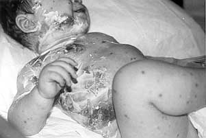
three patients were males. One was 16 years old, the second was an infant whose extensive
skin lesions complicated a viral infection (most probably chickenpox) (Fig. 1), while the
third was an elderly person in whom an allergic reaction to a non-steroidal
anti-inflammatory drug was the most likely causative factor.
|
Fig.1- Infant with post-viral
toxic epidermal necrolysis. |
|
In these two last patients
the diagnosis was readily reached and adequate therapy was swiftly implemented; the skin
lesions healed within the expected time with no or minimal residual scarring. The course
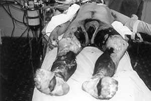
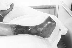
of the first patient was, however, more complicated and the outcome more serious (Figs.
2a,b).
|
|
Fig. 2a
- Young patient with inadequately managed toxic epic necrolvsis resultimi in
full-thickness skin necrosis. |
Fig. 2b -
Same patient after transmetatarsal amputation of foe |
|
The young man was
originally admitted to the medical ward. Despite a dermatological consultation, the proper
diagnosis was not immediately made. The cutaneous lesions were mismanaged and became
desiccated, and the patient also developed ischaemia in both feet secondary to lower
extremity compartment syndrome. Fasciotomy, however, was performed late by the vascular
surgeon. Subsequently, transmetatarsal amputation of one foot had to be carried out.
Postoperatively the patient had to be kept intubated and he was transferred to the
intensive care unit because of severe aspiration pneumonia. It was only then, and after a
previously performed skin biopsy hinted at the possibility of TEN, that the plastic
surgery team was consulted. At that point, all the cutaneous lesions that originally were
salvageable had developed into full-thickness skin necrosis and had to be serially
debrided and skin-grafted. After a long and protracted course, with an episode of cardiac
arrest, the patient gradually recovered and was discharged home. Retrospectively, the
patient's illness was most likely due to anti-epileptic therapy.
Discussion
Adequate diagnosis and prompt
appropriate therapy could have prevented the terrible complications encountered in the
first patient described. rrespective of the exact aetiological factors, the acute,
generalized, sheet-like loss of epidermis described by Lyell to a large extent resembles a
scald burn. Although it is now generally agreed that patients presenting with such a
clinical picture are better managed in a specialized burn unit, some differences do exist
between the "burn-like" syndrome and burn injury itself. These differences
deserve a better understanding as they may have great implications for the type of
management that patients should receive.
Severe thermal or chemical burn injury produces damage to both the epidermal and the
dermal layers. The thickness of the zone of damage is determined by the intensity of the
injurious agent as well as by the duration of exposure, irrespective of the anatomical
peculiarities of the area of skin affected. In patients presenting with TEN,
histologically there is full-thickness epidermal sloughing with the cleavage plane at the
epidermaldermal junction. Vacuolar alterations at the dermalepidermal junction rapidly
progress, forming a subepidermal bulla containing virtually acellular fluid. The separated
epidermis contains individual as well as groups of necrotic keratinocytes that may also be
encountered along the external sheath of the pilosebaceous apparatus"as well as along
the straight dermal duct of the sweat glands. The basement membrane becomes disrupted,
allowing extravasation of all intravascular components. Although a moderate degree of
oedema is present in the papillary dermis, the reticular dermis is normal. 24 Collagen and
reticular fibres are preserved. Regeneration of the epidermis proceeds from the epithelial
remnants of the sweat glands and the hair follicles, similarly to what is observed in
second-degree burns.
It is worth nothing that since in TEN the dermis is preserved and remains completely
viable (unlike second degree burns), less scarring is to be expected following spontaneous
healing, which usually takes 14 days. It is hence crucial during the healing phase to
protect the preserved dermis from infection, desiccation, and subsequent injury U' by
judicious and carefully executed dressing changes. It is logical that surgical tangential
excision is absolutely contraindicated in TEN and that biological dressings and skin
substitutes must be favoured' rather than classical burn dressings that have an inherent
mechanical debriding capacity.
As in burns, silver sulphadiazine cream has been widely used for topical treatment of TEN.
Its use should however be restricted to patients with no previous history of
hypersensitivity to sulphonamides. Pemphigus-like eruptions were described as early as
1942 in association with sulphadiazine therapy. Short courses of corticosteroids early in
the disease have been advocated by some, but their effectiveness has never been
demonstrated. Peters et al. recommended complete avoidance of steroids in the management
of TEN patients.
As in burn injury, TEN causes massive transepidermal fluid loss with associated
electrolyte imbalance proportional to the extent of skin necrosis. The amount of
intravenous fluid resuscitation is however considerably smaller than that given to
patients with a comparable burn injury. This observation needs further investigation. A
probable explanation is that there is less intradermal vascular injury and that the state
of generalized vascular permeability observed early following a burn injury is absent in
TEN. As a corollary, fluid resuscitation formulas and protocols set for burn injury are
not applicable to patients presenting with TEN. Only two-thirds to three-quarters of the
fluids calculated by most standard burn formulas for the first 24 h may be needed.
In contradistinction to the majority of burn injuries, mucosal involvement is a constant
in TEN. Consequently, endotracheal intubation and insertion of nasogastric tubes or Foley
catheters should be avoided if possible for fear of producing more damage to the
structures involved. Endotracheal intubation may traumatize the friable mucosa of the
pharynx, thus contaminating the bronchotracheal tree and precipitating pneumonia.
A peculiarly frequent lesion in patients presenting with TEN, encountered to a lesser
extent also in some face burns, is damage to the cornea associated with severe
photophobia. This type of lesion deserves special attention and extra care that is not
routinely applied to burn patients, if serious ocular complications are to be avoided.
On the other hand, TEN shares with burn injury the necessity to provide the patient with
appropriate nutritional and psychological support as well as adequate narcotic medication
and nursing care in order to minimize suffering, promote non-delayed healing, and prevent
late sequelae and scarring. It is clear that such care can best be provided in a
specialized burn unit.
Conclusion
Although TEN shares many aspects with
burn injury, the peculiarity of the condition deserves special care and attention. TEN
patients should not be managed blindly as burn patients. Health personnel in burn units
should be aware of this fact. Moreover, there is a lot of confusion in the literature
concerning the diagnostic criteria of related conditions presenting with cutaneous
sloughing and necrosis. The argument whether Stevens-Johnson syndrome and TEN are distinct
entities or fall within the spectrum of a single disease process is pointless. Also, the
term "toxic epidermal necrolysis" coined by Lyell in 1956 is a confusing
neologism that does not reflect the exact syndromes may be a better descriptive
classification aetiology of the condition or hint at the way this clinical heading for the
numerous conditions that share in common affliction should be managed. The term burn-like
important features of burn injury.
RESUME. La nécrolyse
épidermique toxique (sigle anglais, TEN) définit un groupe de processus morbides
caractérisés par la lyse mucoépidennique et la nécrose. Cette condition est rare et
peut être associée à une vaste gamme d'étiologies, y inclus la toxicité directe d'un
agent déterminé sur les kératinocytes épidermiques. Dans la forme la plus sévère, le
résultat est normalement funeste. Bien que beaucoup de chercheurs dans ce champ
recommandent pour les patients qui présentent cette condition clinique une gestion du cas
similaire au traitement des patients brûlés, il n'existe pas dans la littérature
scientifique une comparaison approfondie de la maladie des brûlés et de la TEN.
D'ailleurs, le terme TEN, inventé par Lyell en 1956, est un néologisme trompeur qui ne
reflète pas l'étiologie précise de la maladie et ne suggère rien pour ce qui concerne
sa gestion clinique. Puisque la TEN partage avec les brûlures diverses caractéristiques,
dont la plus importante est le traitement recommandé pendant les phases aiguë et
tardives de la maladie, les Auteurs proposent le terme "burn-like syndromes"
("syndromes semblables aux brûlures") pour décrire la vaste gamme de maladies
qui se manifestent avec la formation étendue d'ampoules épidermiques, le dépouillement
de la peau et la nécrose cutanée qui nécessitent l'hospitalisation et une gestion
spécifique dans un service de réanimation.
BIBLIOGRAPHY
- Parsons J.M.: Toxic epidermal necrolysis. Int. J.
Dermatol., 3 t: 749 68, 1992.
- Roujeau J.C., Stern R.S.: Severe cutaneous reactions to
drugs. N. Engl. J. Med., 331: 1272-85, 1994.
- Manders S.M.: Serious and life-threatening drug eruptions.
Am. Family Physician, 51: 1865-72, 1995.
- Peters W., Zaidi J., Douglas L.: Toxic epidermal
necrolysis: a burncentre challenge. Can. Med. Assoc. J., 144: 1477-80, 1991.
- Leape L.L., Brennan T.A., Laird N. et al.: The nature of
adverse events in hospitalized patients: results of the Harvard Medical Practice Study 11.
N. Eng]. J. Med., 324: 377-84. 1991.
- Avakian R., Flowers F.P., Araujo O.E., Ramos-Caro F.A.:
Toxic epidermal necrolysis: a review. J. Am. Acad. Dermatol., 25: 69-79, 1991.
- Chan H.L., Stern R.S., Arndt K.A. et al.: The incidence of
erytherna multiforme, Stevens-Johnson syndrome, and toxic epidermal necrolysis: a
population-based study with particular reference to reactions caused by drugs among
outpatients. Arch. Dermatol., 126: 43-47, 1990.
- Stevens A.M., Johnson F.C.: A new eruptive fever associated
with stomatitis and ophthalmia: report of two cases in children. Am. J. Dis. Child., 24:
526-33, 1922.
- Lyell A.: Toxic epidermal necrolysis: an eruption
resembling scalding of the skin. Br. J. Dermatol, 68: 355-61, 1956.
- Bastuji-Garin S., Rzany B,. Stem R,S. et al.: Clinical
classification of cases of toxic epidermal necrolysis, Stevens-Johnson syndrome, and
erythema multiforme. Arch. Dermatol., 129: 92-6. 1993.
- Roujeau J.C., Guillaume J.C_ Fabre J.P. et al.: Toxic
epidermal necrolysis (Lyell syndrome), incidence and drug etiology in France, 1981-1985.
Arch. Der-matol., 126: 37-42, 1990.
- Goldstein S.M., Wintroub B.W., Elias P.M., Wuepper K.D.:
Toxic epidermal necrolysis. Unnnuddying the waters. Arch. Dermatol., 123: 1153-56, 1987.
- Leenutaphong V., Sivayathorn A., Sithipinittharm P.,
Sunthonpalin P.: Stevens-Johnson syndrome and toxic epidermal necrolysis in Thailand. Int.
J. Dermatol., 32: 428-31, 1993.
- Walker J.: Toxic epidermal necrolysis: theoretical
considerations regarding its aetiology and pathogenesis. Med. Proc., 8: 208-18, 1962.
- Schipf E., Stbluner A, Rzany B. et al.: Toxic epidermal
necrolysis and Stevens-Johnson syndrome, an epidermologic study from West Germany. Arch.
Dermatol., 127: 839-42, 1991.
- Callaway J.L., Tate W.E.: Toxic epidermal necrolysis caused
by "gin and tonic". Arch. Dermatol., 109: 909, 1974.
- Revuz J., Penso D., Roujeau J.C. et al.: Toxic epidermal
necrolysis: clinical findings and prognosis factors in 87 patients. Arch. Dermatol., 123:
1160-5, 1987.
- Villada G., Roujeau J.C., Clerici T. et al.:
ImmuDopathology of toxic epidermal necrolysis: keratinocytes, HLA-DR expression,
Langerhans cells, and mononuclear cells: an immunopathologic study of five cases. Arch.
Dermatol., 128: 50-3, 1992.
- Correcia 0., Delgado L., Ramos J.P. et al.: Cutaneous
T-cell recruitment in toxic epidermal necrolysis: further evidence of CD8+ lymphocyte
involvement. Arch. Dermatol., 129: 466-8, 1993.
- Taylor B., Duffill M.: Toxic epidermal necrolysis from
griseofulvin. J. Am. Acad. Dermatol., 19: 565-7, 1988.
- Amon R.B., Diamond R.L.: Toxic epidermal necrolysis, rapid
differentiation between staphylococcal and drug-induced disease. Arch. Dermatol., Ill:
1433-7, 1975.
- Hurwitz R.M., Rivera H.P., Gooch M.H.: Toxic shock syndrome
or toxic epidermal necrolysis, case reports showing clinical similarity and histologic
separation. J. Am. Acad. Dermatol., 7: 246-54, 1982.
- Massullo R.E., Welton W.A., Jacobsen E.: Toxic epidermal
necrolysis: report of two cases. J. Am. Acad. Dermatol., 19: 359, 1988.
- Solinas A., Cottoni F., Tanda F. et al.: Toxic epidermal
necrolysis in a patient affected by mixed essential cryoglobulinemia. J. Am. Acad.
Dermatol., 18: 1165-9, 1988.
- Eriksson G.: Regeneration of epidermis in Lyell's syndrome,
a light and electron-microscopic study. Acta Dorm. Venereol. (Stockh.), 52: 441-52, 1972.
- Halebian P.H., Shires G.T.: Burn unit treatment of acute
severe exfoliating disorders. Ann. Rev. Med., 40: 137-47, 1989.
- Phillips L.G., Robson M.C., Smith D.J. et a].: Uses and
abuses of a biosynthetic dressing for partial-skin thickness burns. Burns, 15: 254-6,
1989.
- Heimback D.M., Engrav L.H., Marvin J.A. et al.: Toxic
epidermal necrolysis - a step forward in treatment. JAMA, 257: 2171-5, 1987.
- Raffetto J.F., Nichols S.: A nearly fatal reaction to
sulfadiazine in a ten-year-old girl, involving skin, eyes, and oropharynx. J. Pediatr.,
20: 753-5, 1942.
- Sheretz E.F., Jegasothy B.V., Lazarus G.S.: Phenytoin
hypersensitivity reaction presenting with toxic epidermal necrolysis and severe hepatitis:
report of a patient treated with corticosteroid "pulse therapy". J. Am. Acad.
Dermatol., 12: 178-81, 1985.
- Roujeau J.C., Chosidow 0., Saiag P. et al.: Toxic epidermal
necrolysis (Lyell syndrome). J. Am. Acad. Dermatol. 23: 1039-58, 1990.
- Chosidow 0., Delchier J.C., Chaumette M.T. et al.:
Intestinal involvement in drug-induced toxic epidermal necrolysis. Lancet, 337: 928, 1991.
| This paper was received on 10 December 1998. Address correspondence to:
Prof. Bishara S. Adyeh, Division of Plastic and Reconstructive Surgery,
American University of Beirut Medical Centre, Beirut, Lebanon. |
|