Annals of
Burns and Fire Disasters - vol. XII - n° 4 - December 1999
EARLY TANGENTIAL EXCISION WITH THE GUIDANCE OF METHYLENE BLUE APPLICATION
Celikoz B, Deveci M, Nisanci A.
Gulhane Military Medical Academy, Ankara, Turkey
SUMMARY. The difficulty of distinguishing devitalized tissues from
vital tissues in the early post-burn period has long been a deterrent to adequate
debridement. Clinical observation is still the only standard method for estimating what is
to be excised, but it cannot reliably identify the critical zone of vascular stasis that
separates necrotic from viable tissue. In our present clinical study we have evaluated the
reliability of a practical method in which methylene blue is used to predict burn depth.
The mechanism underlying this procedure exploits the metabolism of methylene blue in vital
tissues, where it is converted into leucomethylene by the enzyme reductase. Pre-operative
application of methylene blue enabled us to make an accurate prediction of the depth of
thermal injuries while performing early tangential excision. The mean value of graft take
rates was found to be 98% in all patients treated by skin grafting following tangential
excision performed with the method of pre-operative methylene blue application. Acceptable
wound healing, increased graft survival, and diminished intra-operative blood loss appear
to be the main advantages provided by this procedure compared with conventional early
tangential excision. In conclusion, we highly recommend performing early tangential
excision with the guidance provided by the application of methylene blue.
Introduction
The extent and depth of
thermal injury, the presence of a pre-morbid pathology, and the patient's age are the
primary determinants of mortality and morbidity following thermal injury. Among these,
burn depth deserves special consideration in terms of the patient's long-term appearance
and functioning. In recent years, with better understanding of burn shock pathophysiology,
the resuscitation of burn shock patients is successfully carried out by most surgeons.
However, mortality and morbidity rates remain high with respect to extent and depth of the
burn wound.
Treatment of burn shock and early tangential excision with autografting are the mainstay
of management in deep burns. Inadequate eschar excision leads to skin graft loss, which
results in enlargement of the total area of open wounds with the addition of graft donor
areas and inevitable subsequent additional operations. Although small burns in the hands
and feet are not a life-threatening risk for patients, such wounds have to be treated in
the early post-burn period if patients are to return to their daily and social activities
as soon as possible. First- and second-degree superficial burn wounds in the extremities
are treated by non-surgical methods such as hydrotheraphy, but the treatment of deep burn
wounds, i.e. second- and thirddegree, requires effective surgical treatment techniques.
Appropriate eschar excision in deep burns prevents not only the loss of functional ability
but also the non-cosmetic appearance of the hands, feet, and other parts of the body. The
modem treatment of deep burns therefore entails early surgical excision rather than
waiting for spontaneous eschar separation.
Because of the importance of burn depth in burn treatment, several methods have been
suggested to enhance the accuracy of burn depth prediction. However, despite the advances
of modem technology, there is as yet no method for the precise determination of burn
depth, clinical observation still being the standard method. During conventional
tangential excision, the surgeon considers punctuate capillary bleeding in order to
delineate underlying viable tissue Bleeding points may not be seen during the excision
stage when a tourniquet is used, while blood loss is extensive when the tourniquet is
deflated. It is thus crucial that this aggressive surgical procedure must depend on
accurate recognition of burn depth in order to prevent the unnecessary excision of healthy
tissue and significant blood loss that may jeopardize the patient's chances of survival. A
method that can provide a precise indication of burn depth has thus become a subject of
primary consideration for surgeons.
In our present clinical study, we have evaluated the reliability of a practical method in
which methylene blue is used for predicting burn depth during the early tangential
excision stage. This method consists of the application of methylene blue over the burned
area one day prior to the operation.
Materials and method
Between January 1997 and
March 1998, 42 patients with full-thickness or deep partial-thickness burn injuries were
treated by early tangential excision associated with an autograft procedure in which a new
method of predicting burn depth was employed. Thii~y-one male patients (74%) and eleven
female patients (26%) with a mean age of 24 yr and a mean burn extent of 27% T13SA
underwent early tangential excision between 4 and 7 days post-burn.
Gauze impregnated with a 5% concentration of methylene blue was wrapped over the burned
areas 12 h before the operation (Fig. 1).
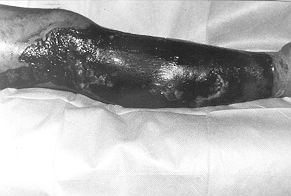 |
Fig.
1 - Appearance of burned leg 12 h after application of methylene blue dye. Notice
the variable coloration of the burn surface in relation to the variable depth of the
thermal injury. |
|
Tourniquets
were applied to prevent bleeding in all the patients subjected to operation. The
blue-stained eschar tissues were excised by deepening the excision until the unstained
tissue border was reached. After completion of the excision at the upper level of the
unstained tissue, the tourniquet was deflated and bleeding was controlled by
electrocautery. It was observed that there was less bleeding, as evidenced by the scarcity
of bleeding points, when this method was compared with conventional excision techniques (Fig.
2).
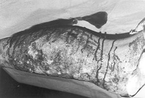 |
Fig.
2 - Same leg after burn wound excision aided by intra-operative application of
methylene blue. Notice the sparse bleeding, less than that usually occurring following
conventional excision performed without the guidance of methylene blue. |
|
Autograft
procedures were performed to cover the excised areas followed by conventional dressing
with vaseline-impregnated gauze. Blood transfusions were not necessary for any of the
patients, owing to the diminished blood loss with our method.
Results
The average percentage of
the full-thickness burn areas treated with the procedure of pre-operative methylene blue
application followed by early tangential excision and auto2raftinR was 23%. The mean graft
take rate was 98% (Fig. 3).
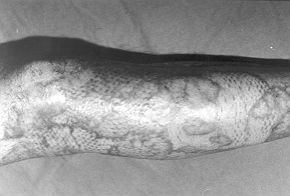 |
Fig.
3 - Final acceptable result after grafting of excised areas. |
|
One patient
underwent secondary skin grafting because of minimal graft loss. No live tissue was
encountered in histopathological studies. The application of methylene blue thus enabled
us to discriminate necrotic tissue from viable tissue.
Discussion
The extent and depth of
burns are essential determinants of mortality and morbidity following thermal injury. A
number of techniques have been defined for the determination of burn depth, such as wound
biopsy, the pin prick test, ultrasound, vital dyes, laser Doppler perfusion monitoring
(LDPM), laser Doppler imaging (LDI) magnetic resonance imaging (MRI), thermal imaging, and
light reflectance or spectroscopic techniques.
Histological dermal biopsy has been reported to diagnose dermal burn depth accurately. One
or more punch biopsies are taken from regions of clinically indeterminate burn depth.
Although histological dermal wound biopsy seems to be the most precise method, the
technique is not without its drawbacks. First, the technique requires an experienced
pathologist to distinguish live collagen from denatured collagen and living cells from
dead cells. Second, since a burn wound continues to have dynamic properties for several
days, wound biopsy is not practical with regard to early tangential excision within the
first 5-7 days of burn injury; it therefore generally takes 7 days to obtain reproducible
results. Third, biopsies leave permanent scars in partial-thickness wounds. Fourth,
biopsies are expensive and time-consuming techniques. For all these reasons, wound biopsy
is rarely used in clinical practice.
B-mode ultrasound scanning was first used by KaluS7 to assess burn depth by observing the
presence of dermal boundaries. Watchel et al. however reported that ultrasonic studies
failed to show burn depth in controlled studies using clinical evaluation and histological
parameters. It was shown that collagen denatures at 65 °C while the epidermal cells from
which burns heal are killed at about 47 °C. Nevertheless, the use of ultrasound in the
determination of burn depth presents some technical difficulties. The requirement of
tissue contact is a further limitation of the ultrasound technique. As a result, apparent
depth is likely to be underestimated by ultrasound.
Laser Doppler or light reflectance has been reported to be effective in the estimation of
full-thickness burn injury. LDPM and LDI have been used for determination of burn depth.
Excellent results were reported in early studies. However, laser Doppler flowmetry, as
also thermography, is highly dependent on certain external conditions, such as room and
body temperature, the patient's anxiety and stress level, and the body area considered.
Laser Doppler flowmetry is non-invasive, easy to use, but expensive, and it has not been
used routinely in clinical applications. It is also time-consuming and too slow for
clinical decision-making.
MR1 depends on the water content of tissues. Koruda et al. reported that in the immediate
post-burn period MR1 could distinguish the higher water content in partial- and
full-thickness rat burns from that in adjacent normal skin. They found that within 48 h
partial-thickness burns returned to control values, while full-thickness burn remained
oedematous. MR1 could therefore be used to distinguish full-thickness burns from
partial-thickness burns. Clinically speaking, however, MR1 is still not very practical.
Vital dyes such as India ink, blue V, toluidine blue, trypan blue, Evans blue, and
disulphine blue have been used to provide sharp demarcation between living and necrotic
tissues. The clinical use of these techniques has however shown that they do not produce
sharp enough demarcation to guide excision Fluorescein flowmetry can be used for the
determination of burn depth since fluorescein, injected systemically, can be detected by
ultraviolet light. It has however been reported by Heimbach et al., Gatti et al., and
Black that the technique can diagnose only very deep and very superficial burns, while the
distinction between intermediate and deep dermal burns cannot not be made, a distinction
that is crucial in operative planning.
In the present study, methylene blue was used to distinguish vital tissue from necrotic
tissue in patients subjected to early tangential excision. Methylene blue is mainly used
to treat methaemoglobinaemia. It has a mildly antiseptic activity and is also used as a
dye in diagnostic procedures such as fistula detection. Methylene blue is metabolized and
converted into leucomethylene by vital tissues following application. Since leucomethylene
is a colourless dye, it is easy to distinguish non-vital tissues from vital tissues, as
described here. During the application procedure, methylene blue impregnated gauze was
wrapped over the burned area 12 h prior to the operation. It can also be mixed with silver
sulphadiazine or other topical burn agents, although impregnated gauze application alone
proves easier than other applications. We observed that at the time of surgery necrotic
tissue was coloured but viable tissue was not. All non-vital tissues were therefore
removed when coloured tissues were completely excised. This procedure helped us to
determine the demarcation zone and minimize vital tissue excision and blood loss.
In conclusion, none of the above-mentioned techniques for the determination of burn depth
is a precise indicator of burn depth at the excision stage, while on the contrary the
application of methylene blue is such an indicator. Pre-operative methylene blue
application enabled us to predict the depth of burn injury accurately while performing
early tangential excision. A precise tissue excision level, appropriate wound healing with
living tissue, increased graft survival, and diminished intraoperative blood loss appear
to be the main advantages of this procedure compared with other techniques. We therefore
highly recommend performing early tangential excision with the guidance provided by the
application of methylene blue.
RESUME. La
difficulté de distinguer entre les tissus dévitalisés depuis longtemps et les tissus
vitaux décourage les chirurgiens d'effectuer un débridement adéquat. L'observation
clinique reste la seule méthode pour évaluer les parties qu'il faut exciser, męme si
elle ne peut pas identifier avec certitude la zone critique de la stase vasculaire qui
sépare le tissu nécrotique du tissu vital. Les Auteurs de cette étude se sont proposés
d'évaluer la fiabilité d'une métode pratique oů le bleu de méthylčne est employé
pour prédire la profondeur de la brűlure. Les mécanismes de cette méthode dépendent
du métabolisme du bleu de méthylčne dans les tissus vitaux oů il est converti en
leucométhylčne par l'enzyme réductase. L'application préopératoire du bleu de
méthylčne a permis aux Auteurs de prédire précisément la profondeur des lésions
thermiques pendant qu'ils effectuaient l'excision précoce tangentielle.
Ils ont trouvé que le taux moyen de prise des greffes était 98% dans tous les pateints
traités avec la greffe cutanée suivie par
l'excision tangentielle effectuée avec l'application préopératoire du bleu de
méthylčne. Les principaux avantages offerts par cette
méthode confrontés avec ceux de l'excision précoce conventionnelle étaient la
guérison acceptable des lésions, la survie augmentée
de la greffe, et la réduction des pertes interopératoires de sang. En conclusion, les
Auteurs recommandent chaleureusement la
technique de l'excision tangentielle précoce avec l'assistance de l'application du bleu
de méthylčne.
BIBLIOGRAPHY
Qelik6z B., Achauer B.M., Vanderkam V.M.:
Hot-press hand treatment. J. Burn Care Rehabil., 19: 128, 1998.
Frist W., Ackroyd F., Burke J., Bondoc C.: Long-term functional result of selective
treatment of hand burns. Am. J. Surg., 149: 76, 1985.
Heimbach D., Mann R., Engrav L.: Evaluation of the burn wound. 16 Management decisions.
In: "Total Burn Care", ed. Herndon D.N., W.B. Saunders, 81-7, 1997.
Hauben D.J., Mahler D.: Histological investigation of burn eschar and underlying
recipient area in tangential early excision of burns. Burns, 5: 160, 1979.
Kahn A.M., McCrady V.L., Rosen V.J.: Burn wound biopsy. Scand. J. Plast. Reconstr.
Surg., 13: 53, 1979.
Laterjet J.: A simple guide to burn treatment. Burns, 21: 221, 1995.
Kalus A.M.: Application of ultrasound in assessing burn depth. 20. Lancet, 28: 188,
1979.
Watchel T.L., Leopald G.R., Frank H.A., Frank D.H.: B-mode 21 ultrasonic echo
determination of depth of thermal injury. Burns, 12: 432, 1986.
Albsjom B., Micheels J., Sorensen B.: Laser Doppler flowmetry measurements of
superficial dermal, deep dermal and subdennal burns. Scand. J. Plast. Reconstr. Surg., 18:
75, 1984.
Essex T.J.H., Byrne P.O.: A laser Doppler scanner for imaging blood flow in skin. J.
Biomed. Eng,, 13: 189, 1991.
Koruda M.J., Zimbler A., Settle R.G.: Assessing burn wound depth using in vitro nuclear
magnetic resonance (NMR). J. Surg. Res., 40: 475, 1986.
Kaufman T., Hurwitz D., Heggers J.: The India ink injection technique to assess the
depth of experimental burn wounds. Burns, 10: 405,1984.
Davies M., Adendorff D., Rode H, van de Reit I.S.: Coloring the damaged tissues on the
burn wound surface. Burns, 6: 156, 1980.
Zawacki B.E., Walker H.L.: An evaluation of patent blue V, brophenol blue, and
tetracycline for the diagnosis of burn depth. Plast. Reconstr. Surg., 54: 459, 1970.
Zuckerman A.: Fluorescein fluorescence photography for the evaluation of burns. J. Biol.
Photogr., 51: 33, 1983.
Gatti J., La Rossa D., Silven-nan D.,
Hantfort C.: Evaluation of the burn wound with perfusion flowmetry. J. Trauma, 23: 202,
1983.
Black K.S., Hewitt C.W., Miller D.M.: Burn
depth evaluation with fluorometry: Is it really definitive? J. Burn Care Rehabil. 7: 313,
1986.
Janzekovich Z.: A new concept in the early excision and grafting of burns. J. Trauma,
10: 1103, 1970.
Jackson D.M.: Historical review of the use of local physical signs in burns. Br. J.
Plast. Surg., 23: 211, 1970.
Jackson D.M., Stone P.A.: Tangential excision and grafting of burns. Br. J. Plast.
Surg., 25: 416, 1972.
Lavrance J.C., Carney S.A.: Tangential
excision of burns; studies on the metabolic activity of the recipient areas for skin
grafts. Br. J. Plast. Surg., 26: 93, 1973.
This paper was received on
3 July 1999.
Address correspondence to:
Dr. Bahattin Celikoz, M.D.,
Docent Dr. G.A.T.A Plastik ve Rekonstrfiktif Cerr. A. Dali
06018 Etlik, Ankara, Turkey.
Tel.: (+90) 312 3045406; fax: (+90) 312 3217778. |
|