Annals of
Burns and Fire Disasters - vol. XIII - n. 2 - June 2000
RESUSCITATION
APPROACH AND CLINICAL EVALUATION IN BURNS VICTIM AFTER MASS ACCIDENTS - PART TWO.
HAEMATOLOGICAL STUDY AND ANALYSES
Hadjiiski O., Rashkov V., Emanuilov J., Lubomirov M.
Centre for Burns and Plastic Surgery,
Pirogov Medical Institute, Sofia, Bulgaria
SUMMARY. This study continues our exploration of the treatment of patients with
burns suffered in mass accidents (Part One was published in the March 2000 issue of Annals
of Burns and Fire Disasters). We observed the changes in various laboratory
parameters, such as the dynamic of the haemoglobin and haematocrit levels, proteins,
potassium, sodium, and the acid-base balance related to the application of Baxter's
modified scheme in the treatment of burn patients in the first three days post-burn. Our
results show a normalization of haematocrit and protein levels, plus a stable electrolyte
balance and satisfactory normalization of the acid-base level
Introduction.
The management of
burns after mass accidents is a frequent problem in Bulgaria, considering the limited size
of our country. Mass burn accidents lead to a great number of deaths and the survivors
usually present a large burn area, which creates treatment problems. Various formulas are
recommended for the stabilization of vital signs after such accidents. We use the modified
Baxter scheme with Ringer's lactate solution in the first 24h after the accident! We
prefer this solution because it is widely known, with clear therapeutic effects, thus
making it very suitable in the event of mass accidents
We include protein solutions after the first 24h, because then the increased capillary
permeability begins to lessen and their deficiency becomes manifest, The studies of the
Cochran Injury Group show that the use of colloids in cases of post-burn hypovolaemia
leads to an the increase,in the mortality rate. Collo ids can be used only in patients
with a pre-existing protein deficit, such as those with inhalation injury, as a
preparation for necrectomy. As a criterion for the effectiveness of treatment we use
various parameters as well as numerous laboratory findings. Some of these are monitored
owing to their doubtful informative value, which on the other hand shows the effectiveness
of the resuscitation efforts. Our results confirm that this scheme of treatment is very
suitable not only in the management of haemodynamic instability but also in the management
of the delicate electrolyte, base-acid and protein balance. Another reason for our
preference for this scheme is the availability of the necessary solutions in every
hospital, as patients are normally transferred early to larger centres where colloids are
available. We use simple parameters for observation, and stabilization continues up to the
end of the period in most patients. The mortality rate is not high. The non-use of blood
transfusion with this scheme means that haemoglobin (Hb) and haematocrit (Hct) values are
not altered.
Materials and methods
The results of the
treatment of 43 burn victims form the object of observation and statistical analysis. We
divided the patients into three groups on the basis of burn area:
- first group - 13 patients, burns up to 29%
TBSA
- second group - 16 patients, burns from 30
to 59% TBSA
- third group - 14 patients, burns from 60 to
100% TBSA
The great majority of the
patients (37) were male; only six were female. The commonest burn agent was flame (32
patients), while scalding accounted for 11 patients. The burn trauma was usually
associated with other injuries. We diagnosed 60 combined injuries (1.40 per patient).
The patients were treated according to the scheme proposed by the team of the SIEM Pirogov
Burns and Plastic Surgery Centre, which is accepted in our country as the preferred scheme
for the treatment of the thermal shock phase. In this study we observed and analysed the
effect of fluid resuscitation on the patients. During thermal shock our scheme of fluid
resuscitation prescribes 3 ml/kg/% TBSA crystalloid (Ringer's lactate) in the first 24 h
period after the accident and 03-0.5 ml/kg/% TBSA protein solutions, plus 1.0-1.5 rnl/kg/%
TBSA 5% glucose solution in the second and third 24 h period after the accident. For more
precise details, Part One of this article will be of assistance.
The following parameters were measured on the first three post-trauma days: Hb, Hct level
at 3-h intervals, levels of base excess from blood gas analysis every 6 h, sodium and
potassium (daily), and plasma proteins.
Results and discussion
Our results show
that the infusions administered were sufficient for stabilization of the general status of
the patients and for their circulation. The patients received a little more than the
calculated quantity of infusion solutions. During the first 24-h period they received only
crystalloids - the quantity received was 10.17 to 10.79% higher than that calculated.
During the following two days, when consolidation of the blood vessel wall began, we
compensated for protein losses - the necessary volume was obtained by means of free water
solutions. The total volume was also higher than that calculated (10.32 to 11.89%), in
spite of the burn area range.
During the second and third 24-h period we used as colloids 5% human serum alburnin or
plasma. The quantity was 0.3 ml/kg/O/o TBSA in patients with burns in up to 29% TBSA,
0.3-0.4 ml/kg/O/o in patients with 30-50% TBSA burns, and 0.4-0.5 ml/kg/% in patients with
over 60% TBSA burns. Patients in the first and second groups received this quantity with a
variation of 6-11%. Patients in the third group received 42-46% less than the quantity
calculated with a significant difference (p < 0.01).
Despite the individual characteristics of each single patient, the proposed quantity and
type of solutions during the period were accurately calculated for all the groups. The
cause of the statistically significant insufficiency of the infusion therapy in patients
with burns in over 60% TBSA on days 2 and 3 can be attributed to the large quantity of
protein solution that we were Lmable to provide in time.
In order to ensure the efficiency of the treatment, we not only followed up the patients'
conditions but also kept a dynamic record of various vital parameters. We observed three
patient groups in order to be able to compare the results in the respective groups and to
provide statistical confirmation.
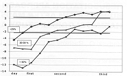
The dynamic of the Hb level is presented in Fig. 1. In the first group of patients,
the Hb level exceeded the normal borderline (159 g/1) only on day 1, while on the
following two days the values were normal. In the second group of patients, the values on
the first two days were over the normal borderline range (184 gft and normalization
occurred on day 3 (151 g/1). In the third group of patients, despite the dynamic decrease
the values remained above the normal borderline range (193 g/l on day 1; 159 g/l at the
end of the period).
The mean values in all the groups were highest on the day of the trauma, as a result of
blood concentration. The results clearly indicate that there was a definite correlation
between the extent of the burn and the timing of the restoration of fluid losses. The
increase in the first group (burns up to 29% TBSA) was 15.2%, in the second it was 32%,
and in the third (burns over 60% TBSA) the increase was 38.2% over the accepted normal
value; the difference between the groups was statistically significant.
In the first group normalization occurred 24 h after initiation of treatment and in the
second group half-way through day 3; in the third group it remained 14.3% above the normal
range after treatment, which shows partial restoration of blood concentration in this
group.
The statistically significant differences at the beginning and the end of treatment show
the reliability of the results in the interpretation of the treatment and the level of
restoration of haemodynamics.
The Hct dynamic is presented in Fig. 2. In the first group of patients the Hct was
above normal values (0.50 ± 0.06) until the 40th hour after the accident, after which it
slowly decreased; at the end day it was normal (0.43 ± 0.07). This finding indicates a
moderate blood concentration on day 1 and in the first half of day 2, followed by
normalization of the circulating blood volume. The statistical analysis as connected with
the different hours excerpt show that the mean Hct values were from 0.53 to 0.42, standard
deviation (SD) from ± 0.07 to ± 0.04, and standard error (SE) from 0.034 to 0.021, where
the significant rate is p < 0.039, proving the reliability of the 250 results.
In the second group of patients, the Het was above the normal borderline (from 0.57 to
0.49 ± 0.07 g/1 at the end of day 2) until the 48th h after the accident, after which it
slowly decreased to normal values on day 3 (0.40 ± 911 0.06). This finding shows a marked
blood concentration on the first two days and gradual normalization of the 100 circulating
blood volume on the third day post-trauma. The statistical analysis of the data shows that
the mean Het values were statistically significant during the whole period in all the
groups compared. The values were from 0.58 to 0.40 gfi (standard deviation from 0.078 to
± 0.049, standard error from 0.017 to 0.022), with a high significance rate of 0.01 to
0.003 (p < 0.01), confirming the reliability of the results.
In the third group of patients, the Het remained above normal borderline values (0.50 ±
0.09) up to the 48th h after the accident and then gradually decreased, until at the end
of day 3 it was below the normal borderline (0.39 ± 0. 12). This finding shows blood
concentration in the first two days and the development of anaemia owing to blood losses
from the wound surface during dressing procedures and to the onset of infection in some
patients, with correspondingly low Hb. The statistical analysis of the data shows that the
mean Het values were statistically significant during the whole period in all the groups
compared, while the significance rate of 0.01 to 0.003 confirms the reliability of the
results.
This parameter is significant for estimating the hypovolaemic state. The mean Het values
were highest on the day of the trauma. The reason for this is the same as for the Hb blood
concentration. The increase in the first group was 21.4% over the normal range, but in
different investigations this difference was statistically non significant (38% in the
second group and 41.67% in the third group above the normal borderline); the differences
between the groups were statistically significant. The Het gradually level decreased but
at the end of the observation period it was near the normal range. In some studies it was
below the normal range; the differences with few exceptions were statistically
significant. There was a definite correlation between burn extent and the time necessary
for restoration of fluid losses.
With regard to the blood acid-base balance, several parameters were observed: blood pH,
oxygen saturation, quantity of carbon dioxide, etc. Fig. 3 illustrates one of these
parameters - the base excess (BE). In the first group of patients BE was minus 4.94 ±
3.59 at the beginning of the period, which confirms the disturbance in the metabolism and
acidosis. However, this parameter normalized at the beginning of the second day. In
patients in the second group, BE was below the normal borderline range (minus 6.87
± 6.7).
This was a
result of dehydration, disturbed tissue metabolism, and the presence of acidosis. Acidosis
decreased on day 2 and around the 60th h BE was positive.
Also in the third group of patients BE was below the normal borderline range (minus 11.85
± 4.52), showing a disturbed tissue metabolism and severe acidosis. The acidosis
decreased on the second day and at the end of the period it was around normal values
(1.40). In the first group of patients treatment of this disturbance was achieved by
correct fluid resuscitation, while in the other two groups buffer correction was needed in
more than half of the patients.
The results show that the metabolism was restored on day 2 in patients in the first two
groups and on day 3 in the third group. The statistically significant difference at the
beginning and end of the period confirms the reliability of the results and the
effectiveness of the treatment.
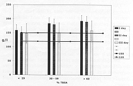
The patients plasma protein dynamic,is presented in Table I. In the first group of
patients the protein value was normal on day 1, and a moderate decrease was noted at the
end of treatment (56.16 g/1). In the other two groups the values were below the normal
borderline range on day 1 and progressively decreased on day 2; at the end of the period
it rose slowly but remained lower than the first value measured. Our observations show
that the changes in plasma proteins were related to the area of the burn and that proteins
reduced as the burn area increased. In the first group of patients the values were near
normal, while in the other two groups the mean values were markedly reduced. In 38% of the
patients in the second group these values were below the normal range. In the third group
the plasma protein loss was especially great, and in 80% of patients it was below the
normal range. The differences in the plasma protein values between the different groups
were statistically significant and were correlated with the burn area and the treatment
effected (insufficient protein replacement in patients in the third group).
| Day |
First |
Second |
Third |
| N |
X |
S |
P |
N |
X |
S |
P |
N |
X |
S |
P |
1-30%
(I) |
10 |
64.9 |
4.95 |
I/II
0.003 |
8 |
7.3 |
6.77 |
I/II
0.01 |
6 |
6.1 |
7.44 |
I/II
0.01 |
31-60%
(II) |
15 |
59.2 |
3.67 |
II/III
0.05 |
13 |
0.4 |
9.7 |
II/III
0.001 |
13 |
4.3 |
7.35 |
II/III
0.002 |
61-100%
(III) |
14 |
48.4 |
17.4 |
I/III
0.001 |
11 |
1.7 |
11.3 |
I/III
0.001 |
13 |
1.0 |
7.16 |
I/III
0.02 |
| Table
I - Dynamic of the plasma protein level of the observed patients |
|
The sodium
dynamic is presented in Table II. In the first group of patients the sodium value
was in the low normal borderline range; the differences in values at the beginning and end
of the period were not significant and did not have a common dynamic. The same holds for
the second group, except that the sodium values were near the upper borderline normal
range. In the third group, where the quantity of the infused solutions was high, the
sodium level dynamic changed. The values were at the upper border of the normal range and
in 25% of group 2 patients and in 31.23% of patients on day 3 the values were above the
normal range. Our results show that in spite of the massive infusion of enriched sodium
solutions, if the quality and quantity of the fluids is correctly calculated there is no
risk of hypernatraernia. The statistical significance of the comparative results in every
group at the beginning and end of the period confirms their reliability and the
correct choice of treatment. The potassium dynamic can be seen in Table III.
| Day |
First |
Second |
Third |
| N |
X |
S |
P |
N |
X |
S |
P |
N |
X |
S |
P |
1-30%
(I) |
11 |
37.1 |
2.3 |
I/II
0.01 |
9 |
36.1 |
5.03 |
I/II
0.01 |
6 |
35.0 |
2.53 |
I/II
0.05 |
31-60%
(II) |
16 |
41.2 |
3.8 |
I/II
0.01 |
13 |
43.1 |
6.76 |
II/III
0.01 |
13 |
41.8 |
10.5 |
II/III
0.001 |
61-100%
(III) |
14 |
45.2 |
4.8 |
I/III
0.01 |
11 |
48.1 |
6.25 |
I/III
0.05 |
13 |
52.9 |
6.62 |
I/III
0.001 |
| Table II - Dynamic of the sodium level of the
patients |
|
| Day |
First |
Second |
Third |
| N |
X |
S |
P |
N |
X |
S |
P |
N |
X |
S |
P |
1-30%
(I) |
11 |
3.89 |
0.45 |
I/II
0.01 |
9 |
4.25 |
0.89 |
I/II
0.1 |
7 |
4.39 |
0.65 |
I/II
0.1 |
31-60%
(II) |
16 |
3.80 |
0.68 |
II/III
0.05 |
13 |
4.26 |
0.99 |
II/II
0.1 |
11 |
4.35 |
0.83 |
II/III
0.1 |
61-100%
(III) |
14 |
4.70 |
1.10 |
I/III
0.05 |
11 |
4.27 |
0.78 |
I/II
0.1 |
9 |
4.26 |
0.70 |
I/III
0.01 |
| Table III - Dynamic of the potassium level of the
patients |
|
The
potassium values show that the mean values remained within the normal range. In the first
and the second groups of patients, these values were lower on the first day and rose
progressively on the other two days, remaining however in the normal range. The
differences between the two groups were not statistically significant. In the third group
the mean plasma potassium values on day 1 were higher than those of the first two groups
and remained the same on the following days. The differences compared with the other
groups were statistically significant. The reason for the higher potassium levels on the
first days was the destruction of cells due to the thermal trauma.
Conclusions
The results of this study,
which is based on findings in 43 victims of mass burn accidents over a 5-yr period, enable
us to recommend the proposed scheme of treatment as a method of choice in the management
of the burn shock phase. The scheme is especially suitable for practical use in mass burn
accidents because of the simplicity and ease of its application, the ready availability in
every hospital of infusion solutions, and the satisfactory nature of the results.
Monitoring includes non-specific laboratory parameters that can be obtained in any
hospital. Normalization of the indices for most patients continues until the end of the
shock phase period.
RESUME. Les
Auteurs confinuent leur étude du traitement des patients brűlés victimes de grands
désastres (la premiére partie a été publiée dans Annals of Burns and Fire
Disasters dans le numéro de mars 2000). Ils observent les modifications des
paramétres de laboratoire, comme la dynamique du niveau de l'hémoglobine et de
l'hématocrite, des protéines, du potassium, du sodium, comme aussi de 1'équilibre
acide-base lié ŕ l'application du méthode modifié de Baxter dans le traitement des
brűlés pendant les premiers trois jours aprés l'accident. Les résultats obtenus
indiquent une normalisation des niveaux de l'hématocrite et des niveaux protéiques,
comme aussi un équilibre stable des électrolytes et une normalisation satisfaisante du
niveau acide-base.
BIBLIOGRAPHY
axter C.R.: Fluid volume and electrolyte
changes of the early postburn period. Surg. Clin. North Am., 58, 1313-22, 1978.
Hadjiiski O.: Therapeutic approach to
emergency disaster situations. J. Emergency Medicine, 6: 71-7, 1998.
Boeckx W.: Fluid resuscitation in mass burn
casualties. International Congress on the Management of Mass Burn Casualties.
Gunn S.W.A., Masellis M.: The World Health
Organization Centre for prevention and treatment of burns and fire disasters: The
Mediterranean Club for burns and Fire Disasters. Ann. burns and Fire Disasters, 11. 3-6,
1998
Cochran injury group: Human alburnin
administration in critically ill patients - systematic review of randomised controlled
trials. Br. Med. J., 25: 235-40, 1998.
Muir I.F., Barclay T.L., Settle J.A.:
"burns and Their Treatment". Butterworth, Oxford, 177, 1987.
Echinard Ch., Latarjet J.: "Les
Brűlures". Masson, Paris, Milan, Barcelona, 349, 1993.
Hadjiiski 0.: Methodological instruction
for action at mass burns accidents. Official Bulletin, Ministry of Health, 6-7: 124, 1996.
Dressler D.P., Hozid U., Nathan P.: Thermal
Injury. C.V. Mosby Co., Washington, Toronto, 93, 1988.
Barton R.G., Saffle J.R., Morris S.E., Mone
M., Davis B., Shelby J.: Resuscitation of thermally injured patients with oxygen transport
criteria as goals of therapy. J. Burn Care Rehabil., IS: 1-9, 1997.
Hadjiiski O., Rashkov V., Emanuilov J.,
Lubomirov M.: Resuscitation approach and clinical evaluation in burn victims after mass
accidents. Part One. Ann. burns and Fire Disasters, 13: 41-5, 2000.
This paper was received on
13 December 1999
Address correspondence to: Prof. Ognian Hadjiiski,
D.MSc., Burns and Plastic Surgery Centre, Pirogov Medical Institute
Blvd Macedonia 21, 1606, Sofia, Bulgaria.
Tel./fax: + 359 2 546108; e-mail: burns_hadj@hotmail.com |
|