Annals of
Burns and Fire Disasters - vol. XX - n. 2 - June 2000
THE
COMPLICATIONS OF BURNS IN THE NEWBORN PERIOD
S. Golubovic Z, Parabucki
D, Janjic G, Zamaklar Cl., Najdanovic Z, Rakic I.
University Children's
Hospital, Belgrade, Yugoslavia
SUMMARY. Over a four-yr period we treated five children
under one month of age for severe burns. All the burns were of iatrogenic origin. The
injury mechanisms were: 1. bathing the child in water that was too hot; 2. use of a hot
water bottle to increase the temperature in the incubator (two cases); 3. use of a hot
water bottle without an incubator; 4. incorrect fitting of the thermocautery electrode to
the child's crura. In all but one of the patients we used the method of tangential
excision after definite demarcation of the necrotic surface. To cover the burn areas we
used homograft skin from the parents and subsequently free skin transplants from the
babies themselves. On the basis of our experience we consider it imperative to treat burns
in neonates following the same principles as in older children. Sepsis was a major
complication in one case but was adequately treated with antibiotics. Later complications
were post-osteomyelitis sequelac and contractures in scars in the groin region.
Introduction
Neonates and, in
particular, premature infants are a high-risk population because of their immature
homeopathic and immune systems. Although burn injury is very rare in this age group it is
important to know how to deal with it. Frequently, even a small lesion - a minor injury
compared with full-thickness skin burns - can lead to sepsis, which is often lethal at
this age.' Sepsis as a complication of burns is the greatest problem, especially if it is
caused by resistant hospital bacteria. The thinness of babies' skin transforms burns that
in older children or adults would cause only superficial injuries into full-thickness
burns.
Materials and methods
Over a four-year period, at the University Children's Hospital in Belgrade, we treated
five babies aged less than one month suffering from severe burn injuries in various parts
of the body. All the injuries were the product of carelessness by the medical staff or
faulty medical equipment, which was not uncommon in past years in our country for
well-known reasons (economic sanctions, war, etc.). All the patients were started on fluid
restitution by peripheral i.v. route, local treatment of the burn area, and parenteral
antibiotics.
Case reports
Patient 1
The youngest baby we treated in our hospital was a 20-day-old premature boy weighing
1700 g. He had sustained 30% TBSA burns during bathing by a nurse in another paediatric
hospital. This occurred because of a faulty water tap that suddenly released hot water
only (Fig. 1). The nurse realised something was wrong the moment water splashed on her
hand. She herself sustained burn injuries on the first digit. The child suffered deep
burns in the flank, back, perineum, and both legs. Initial rehydration was performed in
the same institution and the child was thus not in a bad general condition on arrival. On
admission to our hospital the burn was cleaned, the blisters were removed, and silver
sulphadiazine was applied locally. Continuous parenteral rehydration and i.v. antibiotics
were administered. After four days it became clear that we were dealing with deep burns,
and we therefore we performed tangential excision of necrotic skin and partially covered
the defects with autografts. It was very difficult to harvest autografts because of the
scattered nature of the burns. The rest of the wound was covered with homografts taken
from the patient's father.
Subsequently the child developed sepsis caused by methicillin-resistant Staphylococcus and
arthritis of both glenointmeral joints. An intravenous combination of sulphamethoxasole
and trimethroprim on the basis of the antibiogram, incision, and drainage of both shoulder
joints resolved the sepsis.' All the autografts took, and the parts covered with
homotransplants after reduction healed by second intention. At the age of 1 yr an
operation was performed because of a scar that was pulling the hip into flexion.
The functional results are currently satisfactory, except that osteomyelitis has destroyed
the proximal parts of both humoral bones, causing shortening of length and ankylosis.
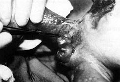 |
| Fig. 1 - Baby with deep burns in flank, back, perineum,
and both legs. |
|
Patients
2 and 3
These two patients are presented together because they sustained burns in the same
incubator at the same time and in the same circumstances. Because of a power-cut, a hot
water bottle was placed between the babies to provide additional warmth. The prolonged
contact of the babies with the bottle caused deep burns. One baby sustained burns in the
brachial and cubital regions, as well as superficial burns in the thigh and scalp (Fig.
2). The other baby sustained burns of the thigh and under-knee. After evaluation of
burn depth tangential excision was performed and the defect was covered in a first
operation with homografts. When the babies' general condition became stabilized the
excised areas were covered with autografts in a second operation. The overall results were
good, and the babies left hospital 14 days after admission.
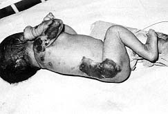 |
| Fig.
2 - Baby with burns in brachial and cubital region and burns of thigh and scalp. |
|
Patient
4
This was the only burn that occurred in our hospital. It was a burn of the under-knee
caused by a faulty thermocautery electrode during surgery for other reasons. Most of the
burn was superficial and only a small part was deep. The burn was treated conservatively.
The superficial part was quickly epithelialized, and the remainder in 10 days. There was
no indication for operative treatment.
Patient 5
This child was transferred from the maternity ward of another hospital with
second-degree contact burns in the face and hand caused by a hot water bottle that was
used as a thermoform to keep the child warm (Fig. 3). The wounds were treated
conservatively with silver sulphadiazine, resulting in good, rapid healing.
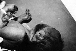 |
| Fig.
3 - Baby with contact burn in face and hand. |
|
Discussion
Burn injury in the
neonatal period is extremely rare, especially in premature babies, and is usually the
fault of medical staff. There have been few reports in the literature about such burns.
They are mainly iatrogenic. The main cause is carelessness and faulty equipment. The
thinness of babies' skin transforrns burns that in adults and older children would be
superficial into deep burns requiring appropriate treatment. Age-related limitations of
the physiological reserves of burned children mean that the adequacy of intravenous fluid
resuscitation is critical. There are few reports in the literature about burns in
premature babies and neonates, which explains the dilemmas posed in treatment. The points
most debated regard which antibiotics and what kinds of local treatment are most
appropriate for use, and whether or not to operate early. The application of 1% silver
sulphadiazine markedly decreases bacterial contamination in the burned surfaces. In our
experience the most important thing is to evaluate each baby singly and to have as much
experience as possible in treating all burns as well as those of surgical neonates.
Our preferred method of treatment is the tangential excision of necrotic skin and the
covering of the defects, preferably with autografts and very often with homografts
whenever the patient's general condition and the presence of scattered burns make
autografts impossible or difficult. Treatment must be rapid because these patients are in
danger of sepsis, even with more banal lesions, and not only with deep and extensive
burns.
RESUME. Les
Auteurs, pendant une période de quatre ans, ont traité cinq enfants d'Age inférieur ŕ
un mois atteints de brűlures graves. Toutes les brillures étaient de nature
iatrogéniqiue. Les causes des brdlures étaient les suivantes: 1. le bain de Fenfant
effectué dans de l'eau trop chaude; 2. la bouillotte utilisée pour augumenter la
température de l'incubateur (deux cas); 3. la bouillotte sans l'emploi de l'incubateur;
4. l'člectrode du thermocautére appliqué en maniére inexacte aux membres inférieurs.
Dans quatre des cinq patients les Auteurs ont utilisé la méthode de l'excision
tangentielle aprés la démarcation de la surface nécrotique. Pour la couverture des
zones brűlures ils ont ernployé l'homogreffe cutanée prélevée des parents, suivie par
des greffes libres cutanées des enfants męmes. Sur la base de leurs résultats les
Auteurs sont convaincus qu'il est n6cessaire de traiter les brűlures des nouveaunés avec
les męmes principes utilisés dans les enfants plus Agés. L'infection a représenté une
complication importante qui a été résolue en maniče efficace avec les antibiotiques.
Les complications successives ont été les séquelles de l'osteomyélite et les
contractures des cicatrices dans la région inguinale.
BIBLIOGRAPHY
Hemdon D.N., Rutan R.L., Rutan T.C.:
Management of the pediatric patient with burns. J. Burn Care Rehabil., 14: 3-8, 1993.
Lochbuhler H., Meuli M.: Current concepts
in pediatric burn care: Surgery of severe burns. Eur. J. Pediatr. Surg., 2: 201-4, 1992.
McAllister R.M., Mercer N.S., Morgan B.D.,
Sanders R.: Early diagnosis of staphylococcal toxacmia in burned children. Burns, 19:
22-25, 1993.
Alison W.E., Jr, Moore M.L., Reilly D.A.,
Phillips L.G., McCauley R.L., Robson M.C.: Reconstruction of foot burn contractures in
children. J. Burn Care Rehabil., 14: 34-38, 1993.
Graves T.A., Cioffl W.G., McManus W.F.,
Mason A.D., Jr, Pruitt B.A., Jr: Fluid resuscitation of infants and children with massive
thermal injury. J. Trauma, 28: 1656-9, 1988.
Kudlackova M.: Antibacterial creams for the
treatment of burns in infants and toddlers. Acta Chit. Plast., 30: 39-43, 1988.
| This paper was received on
3 January 2000. Address
correspondence to: Dr Zoran Golubovic MD, Phd,
Paediatric Surgeon, University Children's Hospital
Tirsova 10, 11000 Belgrade, Yugoslavia. |
|