Annals of
Burns and Fire Disasters - vol. XIII - n. 4 - December 2000
ENHANCEMENT OF BURN HEALING BY GROWTH FACTORS AND IL-8
Blumenfeld I.,1 Ullmann Y.,2
Laufer D.,1 Livne E.3
Faculty of Medicine,
Technion-Israel Institute of Technology, Haifa, Israel
1 Maxillofacial and Oral Surgery Dept., Rambam Medical Centre
2 Plastic Surgery Dept, Rambam Medical Centre
3 Division of Morphological Sciences
SUMMARY. The aim of the present study was to evaluate the
ability of interleukin-8 (IL-8) to enhance wound healing and to compare this with the
effect of transforming growth factor-(31 (TGF-(31) + basic fibroblast growth factor (bFGF)
applied in combination. Inflicted burn injury was used as the model for wound healing.
Deep partial-thickness burns were inflicted using aluminium templates. IL-8 and growth
factors were applied topically using measured volumes (40 pd) and the wound was covered by
non-adherent absorbent dressings and then bandaged. Treatment was repeated every two days
for up to 13 days. Wound areas were recorded and photographed and tissue biopsies were
obtained on the last day for general morphology. The results indicated that treatment with
IL-8 enhanced epithelialization and reduced contraction and the open area values of
inflicted wounds. IL-8 appeared to be the most significant in this respect compared with
TGF-(31 + bFGF or control groups. Morphology obtained from tissue biopsies on the last day
of the experiment also revealed that re-epithelialization was most significant in IL-8
treated wounds. It is concluded that the topical application of IL-8 enhanced the wound
healing process in experimental animals
Introduction
Wound healing is a
complex programmed sequence of cellular and molecular processes including inflammation,
cell migration, angiogenesis, provisional matrix synthesis, collagen deposition, and
re-epithelialization.' Until recently, no pharmacological agents that could reproducibly
accelerate wound healing had been identified. The identification of cytokines and growth
factors as mediators of many of the processes integral to tissue repair has sparked
renewed interest in the development of potential therapeutics to enhance deficient tissue
repair. 2 Growth factors and cytokines are considered candidate therapeutics because they
are synthesized by and stimulate cells required for tissue repair (e.g., platelets,
macrophages, endothelial cells, keratinocytes, and fibroblasts). They are deficient in
chronic wounds, and pharmacological applications to wounds enhance wound repair.
Normal wound healing is thought to occur in three stages: a. directed and sequential
migration of neutrophils, monocytes, keratinocytes, and fibroblasts into the wound over
the first several days; b. activation of wound macrophages and fibroblasts resulting in
the de novo synthesis of growth factors and other cytokines, extracellular matrix (ECM)
proteins and proliferation of fibrobtasts in the successive 2-3 weeks; c. remodelling with
active collagen turnover and crosslinking from two weeks to one year postwounding
.Cytokines are usually grouped into "families" on the basis of structural and
functional singularities. The accumutation and activation of leukocytes in wounded tissues
is the most characteristic expression of infection. The mechanism of leukocyte recruitment
has been studied for decades, and the breakthrough came a few years ago with the discovery
of interleukin-8 (IL-8) and several related chemotactic cytokines. IL-8 and related
chemotactic cytokines are now collectively called chemokines. These are small proteins of
70-80 amino acids with four conserved cysteines that form two disulphide bonds, a short
amino terminal, and a longer carboxyl terminal sequence.' Two subfamilies are
distinguished according to the arrangement of the first two cysteines, which are either
separated by one amino acid (CXC chemokines) or adjacent (CC chemokines). CXC chemokines
act mainly on neutrophil leukocytes while CC chemokines are inactive on these cells.
Cytokines and growth factors have the potential to improve wound healing through several
mechanisms:
- they have chemotactic activities that
attract inflammatory cells, fibroblasts and keratinocytes into the wound;
- they act as mitogens to stimulate cellular
proliferation;
- cytokines and growth factors can stimulate
angiogenesis, the ingrowth of new blood vessels into the wound;
- they have a profound effect on the
production and degradation of the ECM;
- they influence the synthesis of other
cytokines and growth factors by neighbouring cells.
TGF-▀1 isolated from
blood platelets has a major effect on tissues of mesenchymal origin. It has been shown to
induce wound repair following application in rats' and in humans.' Addition of TGF-(3
prior to experimentally induced excision wounds enhanced wound repair.
Fibroblast growth factor (FGF) was also found to induce angiogenesis and stimulate
endothelial and smooth muscle cells.' It was reported that application of bFGF on pressure
wounds in paraplegics led to dose-response healing of the wound.
Growth factors are good candidates for the treatment of wound healing because they are
naturally produced by the cells and enhance tissue repair." Their use in chronic
wounds was found to enhance repair. Clinical studies have shown that FGF applied to
chronic and diabetic ulcers significantly enhanced their repair.
The use of cytokines to enhance wound healing is crucial in cases of burns, chronic
pressure wounds, diabetes wounds, and chronic ulcers. The possibility of enhancing wound
healing by cytokines is beneficial in long-term hospitalization, in the elderly, in
accident wounds, and in burns sustained on the battlefield.
Other studies have been conducted on growth factors, but the possible effect of the
induction of wound repair by IL-8 has not yet been studied. The purpose of the present
research was therefore to study possible ways of enhancing wound injury repair by using
the cytokine IL-8 and to compare the effect of combined treatment with growth factors
(TGF-(3 and bFGF).
Materials and methods
Deep partial-thickness
skin burns were inflicted using guinea-pigs weighing 250-300 g. The animals were
anaesthetized (ketamine HCl, 150 mg/kg i.m.) 24 h prior to the experiment. The back and
abdomen were clipped using electrical clippers (Oster-Golden A-S, model 5-55E, 35 W, head
no. 80, blade size 40). The back and right flank of each animal were then depilated using
a standard depilatory cream. This approach ensured thorough and uniform removal of the
animals' fur. Since clipping and depilation cause oedema of the skin, a period of 24 h was
allowed to elapse before the burn was inflicted.
The thermal source for the burn injury was a cylindrical aluminium template (diameter,
3.76 cm; length of handle, 24 cm; total weight, 500 g). '5 The templates were heated in a
water bath for 2 h prior to the injury at a constant temperature of 75 ░C. Six templates
were heated concurrently and used alternately, one for each injury, and then returned to
the heating water to ensure maintenance of the desired temperature of the template
surface. About 5 min should elapse between re-usages of the same template.
In order to achieve a significant reduction in the biological variables among the various
treated groups of burns in the experiment, the animals were anaesthetized again on the
following day. Prior to infliction of the burn wound, the animals were restrained and
stretched flat on a wire mesh and the midline corresponding to the spine was marked. At
the level of the mid-distance between the twelfth rib and a horizontal line between both
sacroiliac joints on the right-hand side, a point was determined as the exact location of
the injury for all animals.
The heated and moistened template was applied at right angles to the skin of the animal's
back according to the pre-marked location, while monitoring lasted exactly 5 sec, using an
analogue stopwatch (Fisher Houer, Switzerland). Only minimal pressure was required in
order to ensure perfect contact between the template surface and the underlying dorsal
skin.
The animals were divided into three groups (one control group and two experimental). The
experimental animals were treated topically with measured volumes (40 ~d) of IL-8 (25
ng/pl in saline) or with 20 p1 of TGF-(31 (10 ng/pl, dissolved in 4 mM HCl containing 0.1%
BSA) + 20 Id FGF (25 ng/~l in saline), all from Sigma, St Louis, Mo, USA. Control groups
were treated similarly using 40 l-1 of saline.
Following application of cytokine, growth factors, or saline, the wounds were covered with
a non-adherent absorbent dressing (Melolin, Smith and Nephew Ltd, Hull, UK) and bandaged
using elastic adhesive bandages (Salvaplast Ind. Ltd, Petach-Tiqua, Israel).
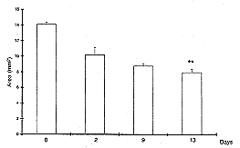
The treatment was repeated every 2 days for up to 13 days. On days 0, 2, 9, and 13 the
diameter of each burn injury was recorded using overhead viewgraph clear transparencies
(one per animal). Burn injuries were also photographed under fixed lens distance
conditions. Upon termination of the experiment the animals were anaesthetized as described
above, and tissue biopsies for light microscopy were obtained from a predetermined area.
Tissue samples designated for light microscopy were fixed in 4% phosphate neutral buffered
formalin, pH 7.4, for 48 h, dehydrated in graded alcohols and embedded in paraplast.
6-um-thick sections were stained with haematoxylin and eosin for general morphology and
photographed using an Olympus CH-2 microscope.
The recorded values of burn injury in each time interval were used to calculate the actual
area (cmz) of "open" and "total" areas of each burn injury on days 0,
2, 9, and 13, using a morphomat system (Morphomat 30 and Epson printer, C. Zeiss,
Oberkochen, Germany). In addition, the values of the "total" area in each
interval, the values of the "open" area on day 13, the "percent of open
area" from the total area on day 0, the "percent of contraction" (total
area on day 0 minus total area on day 13), the values of "epithelialization"
(total area on day 13 minus "open" area on day 13), "epithelia] ization +
"contraction", and percentage "repair" were calculated statistically
using Instat2. Student's t-test and analysis of variance (ANOVA) were performed
(significance level, p < 0.05).
Results
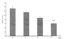
The "total"
area of all injuries appeared to decrease gradually from day 0 up to day 13. However,
compared with the control group (Fig. 1), this reduction appeared to be more
significant (p < 0.001) in burns treated with TGF(P + FGF (Fig. 2) as also in
burns treated with IL-8 (p < 0.001) (Fig.3). This reduction in the
"total" area represented a "contraction" process calculated as
"percent" from the area on day 0 (Fig. 4).
 |
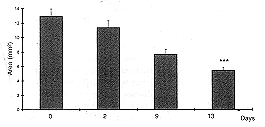 |
| Fig. 1 -
Wound area (cm▓) in control animals (** p < 0.01). |
Fig. 2 -
Wound area Ian=i in animuh treated with TGF-1 + bFGF i *** p < 0.01 i. |
|
|
 |
| Fig. 3 -
Wound area 1 cm=) in animals treated with IL-8 I `*' 1) < 0.001 i. 8 treated animals. |
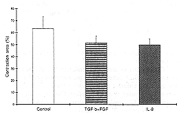
Fig. 4
- Percent contraction in control. TGF-1 1 bFGF and 1L-S heated animals. |
|
It appeared
that "contraction" values following treatment with IL-8. as also after treatment
with TGF-r) + FGF.
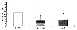
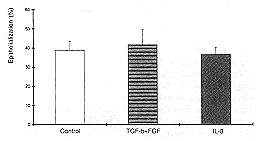
Similarly, the percent values of "open" area of wounds on day 1 3 were reduced
significantly (p < 0.05 ) in both IL-8 and TGF-r) + FGF treated injuries (Fig. 5) compared
with control. Percent values of the "closed" ("epithelialization")
area appeared to be similar in both treated groups compared "-ith control (Fib.
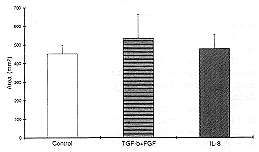
6). The values of "contraction + epithe-lialization" appeared to be higher
in IL-8 as well as in TGF-(3 + FGF treated groups compared with control (Fig. 7). Percent
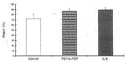
"repair" was higher in the IL-8 treated group than in the TGF-(3 + FGF or
control groups (Fig. 8).
|
 |
| Fig.
5 - Percent open area in control. TGF-1 + bFGF and IL-8 treated animals (* p <
0.05). |
Fig.
6 - Percent epithelialization in control. |
|
|
 |
| Fig. 7 -
Epithelialization + contraction area in TGF-1 + bFGF and IL-8 treated animals. |
Fig. 8 -
Percent of repair in TGF-1 + bFGF and IL-8 treated animals. |
|
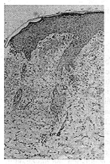
Tissue
morphology revealed that on day 13 a very thin layer of epidermis was visible in the
control group (Fig. 9).
A similar result was obtained in TGF-▀ + FGF treated animals (Fig. 10).
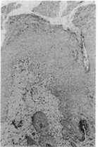
In animals treated with IL-8, thick epidermis as also well-developed skin appendages were
observed (Fig. 11).
Discussion
Cytokines and growth
factors can directly regulate wound healing by affecting the chemotactic attraction of
inflammatory cells, mitosis of fibroblasts, keratinocytes, and endothelial cells as well
as by neovascularization and ECM synthesis.
It is possible that during the process of wound healing the amount and concentration of
certain cytokines and growth factors levels are low or missing, which could slow down or
inhibit the healing process. In chronic pressure wounds, diabetes, or ulcer wounds the
levels of some growth factors were diminished or absent, resulting in slow or non-existent
repair of chronic wounds. No treatment for the repair of such chronic wounds has ever been
suggested.
In the past, various topical agents and bandages were used, but topical treatment with
IL-8 has not been studied previously. The present study tested the effect of IL-8,
demonstrating that topical administration of IL-8 enhanced wound healing. Our innovation
will contribute to the improvement of unhealed wounds in burns, infections, and pressure
wounds and in cases of systemic diseases, diabetes, and insufficient blood supply in
altered angiogenesis.
The criteria used for evaluating wound healing were the measurements of the total area of
the wound at the beginning and on the last day of the experiment, the values of
contraction, the open area of the wound on day 13, the percent of the open area compared
with the total area on day 0, the percent of contraction, epithelialization values, and
percent repair. Our results indicated that the effect of IL-8 was most beneficial with
regard to all criteria tested.
Large burns are followed by significant trauma-induced immunomodulation. The mean burned
patient plasma concentration of IL-8 is about 60 times higher than that of healthy
persons. Also, patients with total body surface area burns had significantly higher IL-8
concentrations in plasma than patients with smaller burns. IL-8 is localized in the basal
germinative cell layer or at the focal sites of ongoing neutrophil inflammation in the
suprabasal cell layer. IL-8 has been implicated as a regulator of the proliferation and
differentiation of normal keratinocytes and as a mediator of keratinocyte maturation and
migration in inflammatory processes involving the skin.
IL-8 may play an important role in the homeostasis of normal epidermis as well as in wound
healing, in which dermal-epidermal interactions and keratinocyte migration are critical.
Dermal factors influence epidermal differentiation and appendage formation, but the rules
governing these interactions and the factors that affect them are poorly understood.
Progressive skin necrosis after trauma such as burn wounds and local trauma is a frequent
occurrence. During this process, tissue initially appears to be viable, with clinical
evidence of perfusion. This tissue eventually dies, which has a profound clinical
significance as the ultimate tissue loss is much greater than that estimated at the time
of initial injury. The injured skin directly initiates an inflammatory response that
includes the release of neutrophil chemoattractants. IL-8 is a specific cytokine initiator
of post-traumatic cascade wound healing. Previously documented properties of IL-8 support
the proposal of this cytokine as an important mediator of wound healing. IL-8 is
synthesized by resident skin cells, e.g., keratinocytes," fibroblasts,"
macrophages, and endothelial cells.
Rot et al. demonstrated the presence of binding sites for IL-8 on the endothelium of
post-capillary venules and small veins in human skin." Another aspect of IL-8 is its
ability to trigger angiogenesis in vivo by indirect action, probably through leukocyte
recruitment."
Also, IL-8 induces increased microvascular permeability in human skin.
IL-8 stimulates neutrophils to degranulate, thereby exposing surface receptors which may
promote integrinmediated adhesion between the neutrophils and the endothelial cells.
The purpose of this study was to test the capacity of IL-8 to enhance wound healing by
topical application. We proved that IL-8 significantly reduced the total injured area more
than the growth factors and the control group. This was to be expected as we are aware of
IL-8's ability to influence and induce dermal fibroblasts and keratinocytes to proliferate
and synthesize. Another aspect of our study which may be promising for the use of IL-8 in
wound healing is the fact that IL-8 reduced wound contraction compared with the control
group. On the other hand, the capacity of IL-8 to regulate the proliferation and
differentiation of keratinocytes in addition to their maturation and migration resulted in
a markedly thick epithelialization layer of the wound area compared with both the control
and the growth factor treated groups.
In the light of the promising and encouraging results of our study, we are optimistic as
regards the power of our innovation to contribute to the improvement and enhancement of
problematic wounds, burn infections, and decubitus problems and of wounds secondary to
systemic diseases such as diabetes, insufficient blood supply, and altered angiogenesis.
To our best knowledge there are no recognized limitations to this treatment. However,
further large-scale experiments are needed before clinical trials can be performed. In
addition, more studies are needed in order to understand the mechanism of this enhancement
of wound healing by IL-8.
RESUME. Dans
cette etude les Auteurs se proposent d'Úvaluer la capacitÚ de 1'interleukine-8 (1L-8)
d'amÚliorer la guÚrison des lesions et d'effectuer une comparaison avec 1'effet du
facteur de croissance transformant-P1 (TGF-P1) + facteur de croissance des fibroblastes
basique (bFGF) appliquÚs en combination. Coinme modÞle de guerison des lesions ont Úte
usÚes des br·lures infligÚes. Des brGlures profondes d'Úpaisseur partielle ont ÚtÚ
effectuÚes utilisant des matrices d'aluminium. L'IL-8 et les facteurs de croissance ont
ÚtÚ appliquÚs topiquement avec 1'cmploi de volumes mesurÚs (40 pl) et de pansements
absorbants non-adherents. Le traitement a Útd rÚpÚtÚ tons les deux jours jusqu'Ó 13
jours. Les zones des lesions ont ÚtÚ notÚes et photographiÚes et des biopsies
tissulaires ont ÚtÚ effectuÚes le dernier jour pour la morphologie gÚnÚrale. Les
rÚsultats indiquent que le traitement avec IL-8 amÚliorait 1'epithdlialisation et
rÚduisait la contraction et les valeurs des zones ouvertes des lesions infligÚes. A cet
dgard 1'IL-8 se prÚsentait comme le plus significatif par rapport Ó TGF-(31 + bFGF on
les groupes tÚmoins. Les examens rnorphologiques effectuÚs avec les biopsies tissulaires
le dernier jour de 1'expÚrimentation ont rÚvÚlÚ que la rÚÚpithÚlialisation Útait
la plus significative dans les lesions traitÚes avec IL-8. Les Auteurs concluent que
1'application topique de 1'1L-8 amÚliorait les processes de la guÚrison des lesions dans
les animaux expÚrimentaux.
BIBLIOGRAPHY
Pierce G.F., Mustoe T.A.: Phatnlacoloeical
enhancement of wound healing. Ann. Rev. Med.. -16: 467-81. 1996.
Greenhalah G..: The role of growth factors
in wound healing. J. Trauma. 41: 159-67. 1996.
Clark R.A.: Basics of Cutaneous Wound
repair. J. Dermatol. Surg. Oncol.. 19: 69 3- 7 06. 199 3.
Clark-Lewis et al.: Structure-activity
relationships of IL-8. J. Biol. 16. Chern- 266: 28-13-1, 1991.
Baff2hioni NI.. Dewald B.- Moser B.:
Interleukin-8 and related chemotactic evtokines-CXC and CC chemokines. Adv. Immun.. 55:
97-179. 1994
Mustoe T.A.. Pierce G.F. Thomson P.D. et
al.: Accelerated healing of incisional Wound in rats induced b_v transforming growth
factorb. Science. 3.17: 1333-6. 1987.
Cromack D.T Poras-Reves B Pordy J. et al.:
Acceleration of 18. tissue repair by transforming growth factor-bl: Identification of in
vivo mechanism of action w ith radiotherapy-induced specific healing deficits.
Surgery. 113: 36--1_'. 1993.
Beck L.S.. De Guzman L.. Lee `W.P. et al.:
One systemic administration of transforming growth factor-b 1 reverses systemic or
alueocorticoid-impaired wound healing. J. Clin. Invest.. 93: 2841-9,1993
Maciaf T.. Zahn h.. Garfinkel S. et al.:
Novel mechanism of fibroblast growth factor function. Rec. Prog. Honn. Res.. 49: 105_'3.
1994
Montesano R., Vassalli J.D.. Baird A. et
al.: Basic fibroblast growth factor induces ansiogenesis in ritro. Proc. \att.
.acad. Sci USA. 8 3: 729 7-301. 1986.
Mellin T.N.. Mennie R.J.. Chashen D.E. et
al.: Acidic fibroblast growth factor accelerates dermal wound healing. Growth Factors, 7:
1-14, 1992.
Cooper D.\'L. I"u Z'.Z., Hennessey P.
et al.: Determination of endogenous c\tokines in chronic wounds. Am. Sura.. 219: 688-92.
1994.
lustoe T.A.. Pierce G.F.. \Iorishima C..
Deuel T.F.: Grow h factor induced acceleration of tissue repair through direct and
inductive activities. J. Clin. Invest.. 87: 694-703, 1991.
Bennett N.T.. Schultz G.S.: Growth factors
and wound healing: Biochemical properties of growth factors and their receptors. Am. J.
Surs.. 165: 7'8-37. 1993.
Kaufman T.. Lusthaus N.. Saghner L.. "
enter bLR.: Deep partial skin thickness burns: ? reproducible animal model to study burn
wound healing. Burns. 16: 1 3-16. 1990.
Fincham N.D., Camps R.D.R.. Gearing A.J.H..
Brid C.R.. Cunningham F.LL: Neutrophil chemoattractant and IL-1-like activity in samples
from psoriatic skin lesions. J. Demrtatol., 140: 294-305. 1988.
Schroder J.__\'1.. Stricherlina M..
Henneicke H.H.. Priessner \.C.. Christopher E.: IL-1a or TNF-a stimulate release of three
'_s1AFI;'IL-B-related neutrophil chemotactic proteins in human dermal fibroblasts, J.
bnmunol.. 1-t4: 2d?3-3'. 1990.
Rot A.. Hub E.. Middleton J.. Pons F..
Rabeck C.. Thierek. V'inttle J.. V olff B.. Zoak NL. Dukor P.: Some aspects of IL-8
pathophy•sioloav. III. Chemokine interactions with endothelial cells. J. Leukoc.
Biol.. 59: 39--1.1. 1996.
Petzelbauer P.. " atson C.A.. Pfau
S.E.. Pober J.S.: IL-8 and anaioQenesis. Cwokine. ?: 26¯-7'. 1995.
Douglass J.. Dhami P.. Bolpitt __\l..
Lindley J.. Chuete J.. Church 1LK.. Hoegate S.T.: Inuadermal challenge with IL-8 causes
tissue oedema and neutrophil accumulation in atopic and non-atopic human subject. Clin.
Eap. -allera~-. _'6: 1371-79. 1996.
This paper was received
on 1 June 2000.
Address correspondence to:
Dr Erella Livne. D.Sc.. Anatomy
and Cell Bioloev.
Faculty of Medicine. Technion. PO Boy 9619. Haifa 31096. Israel
Itel.: 97? -l 8295391: fat: 972 4 82953921. |
|