Annals of
Burns and Fire Disasters - vol. XIV - n.1 - March 2001
USE OF GLUCOCORTICOIDS IN A BURN PATIENT
WITH ADULT RESPIRATORY SYNDROME
Ramos G., Patiho O., Sanchez
Luceros D., Bolgiani A., Brunoldi D.,Prezzavento C., Benaim F.
CEPAQ, Benaim Foundation, Aleman Hospital,
Buenos Aires, Argentina
SUMMARY. We
present a male patient with burns in 23% body surface area. He developed adult respiratory
distress syndrome (AF;DS) and septic shock during evolution. Although infection was under
control, respiratory failure and vasopressor agent requirements did not improve until
glucocorticoid treatment was initiated. Glucocorticoid treatment allowed us to take the
patient off mechanical ventilation and suspend vasopressor agents during the first week of
treatment. The use of glucocorticoids in burn patients, whether suffering from ARDS or
septic shock, is controversial. However, it may be effective in the fibroproliferative
phase of ARDS and septic shock with infection under control, as in the case presented.
Introduction
Septic shock and adult
respiratory distress syndrome (ARDS) are important causes of death in burn patients. These
conditions produce an unstable state between the causative agent and the inflammatory
response, resulting in a mortality rate of approximately 50%.
Sepsis is defined as the systemic inflammatory response to an infection.'
Septic shock is defined as sepsis with hypotension, despite adequate fluid resuscitation,
together with the presence of perfusion abnormalities that may include, but are not
limited to, lactic acidosis, oliguria, or an acute alteration in mental status. Patients
who are on inotropic or vasopressor agents may not be hypotensive at the moment when
perfusion abnormalities are measured.' Septic shock occurs in 20-40% of septic patients,
with a mortality rate of 40-90%. ARDS is defined as acute respiratory failure, with
diffuse pulmonary infiltrates, a Pa02/FiO2 ratio of less than 200 with a pulmonary wedge
pressure (pulmonary artery occlusive pressure) less than 18 cm H20, and absence of
evidence of cardiac pump failure.The causes of ARDS are either pulmonary or extra-
pulmonary.
In burn patients, the most frequent cause of pulmonary dysfunction is inhalation injury,'
while in patients without inhalation injury, ARDS develops as a result of sepsis usually
originating from wound infection. Respiratory injury worsens the prognosis in burn
patients, independently of the severity of the burn injury. ARDS is characterized by a
diffuse alveolar inflammation and increased pulmonary capillary permeability to fluids,
whatever the cardiac pre-load volume may be.There are three phases of ARDS:' the exudative
phase, the fibroproliferative phase, and the pulmonary fibrosis phase. The first of these
phases is characterized by abnormal increases in pulmonary capillary permeability and the
damage caused by oxidants and proteolytic enzymes liberated by inflammatory cells. In some
patients, ARDS resolves itself at this point, while in others it continues until a
fibroproliferative period which prolongs mechanical ventilatory dependence and its related
complications, thus increasing mortality. It is known that basal membrane integrity and
the capacity of Type H pneumocytes to replicate is fundamental to normal healing and the
resolution of ARDS. However, the capacity to recover is dependent not only on the initial
pulmonary lesion but also on subsequent and repetitive conditions such as those secondary
to persistent endotoxaemia, nosocomial infection, the toxic effects of oxygen therapy, and
barotrauma. These complications affect the balance between the reparative and the lytic
processes.
The fibroproliferative phase is characterized by the proliferation of myofibroblasts and
the deposition of collagen, transforming the initial exudative phase into granulation
tissue. Unfortunately, this healing process is inefficient and causes malfunction of the
ventilatory system owing to pulmonary restriction, which characterizes the fibrotic phase
of ARDS.
After the initial injury, the body responds with the activation of an inflammatory cascade
of the coagulation system, the immune system, tissue repair, and the activation of the
hypothalamo-hypophyso-suprarenal system, producing glucocorticoids. The activation of this
last system moderates the response with the result that it does not get worse than the
initial injury. Various methods have been used to treat this inflammatory response, but
without any satisfactory results. Glucocorticoids have been considered not to be
beneficial, and they are potentially deleterious in septic shock and ARDS,"'
especially in burn patients.However, recent studies have demonstrated the utility of
glucocorticoids in the late stages of septic shock and ARDS."'
The objective of this report is to define the role of glucocorticoids during the
intermediate, or fibroproliferative, phase of ARDS, citing the relevant literature.
Case report
We admitted a 26-year-old
male patient with a direct flame burn and a prior history of schizophrenia. The burn
affected 23% of the body surface area, of which 16% was partial thickness and 7% full
thickness. There was no inhalation injury. The burns were located in the face, the
anterior trunk and abdomen, the right arm, and the bilateral upper thighs. Silver
sulphadiazine was applied to the wounds and reapplied every 12 h. On day post-burn 6, the
patient was febrile and presented the clinical criteria of burn wound infection." Proteus
mirabilis and P. aeruginosa, sensitive to ciprofioxacin and amikacin, were
isolated. No other infectious loci were identified. The wounds were excised tangentially
and covered with silver sulphadiazine dressings. On day 8 post-burn, the patient continued
to be febrile and developed episodes of hypotension, accompanied by oliguria, tachycardia,
tachypnoea, disorientation, and hallucinations. Two litres of crystalloid were rapidly
infused without any response, and dopamine infusion was therefore initiated at a rate of
10 mcg/kg/min. Additionally, supplemental oxygen was initiated with aggressive respiratory
therapy, because of a deterioration in gas exchange (OZ saturation fell to less than 90%
of room air). Diffuse pulmonary infiltrates were noted on chest X-ray, and arterial blood
gases were: pH, 7.43; PaC02, 29; Pa02, 83; bicarbonate, 22; 02 saturation, 96.9% on 35%
Fi02 by mask. On day 9 post-burn, a second tangential excision of suspicious wound
infections was performed. Twelve hours post-operatively, the patient was orotracheally
intubated and mechanical ventilation (MV) was initiated following a deterioration of
mental status and further oxygen desaturation. Initially, the patient required 100% Fi02
with a PEEP of 5 cm H20 to produce the following ABG: pH, 7.2; PaC02,46; Pa02, 49; BE,
-10; 02 saturation, 73%. Additionally, the patient became haemodynamically unstable, with
hypotension refractory to fluid expansion and little response to dopamine titration.
Haemodynamic stability was achieved with epinephrine (0.9 mcg/kg/min). A Swan Ganz
catheter was inserted, revealing a PAOP of 8 mm Hg and a cardiac output of 12 1/min. The
following days the patient remained in a hyperdynamic state with a Pa02/Fi02 ratio between
150 and 200. On day 10 (MV day 2), fiberoptic bronchoscopy and bronchoalveolar lavage
demonstrated a significant number of gram-positive cocci and gram-negative bacilli, for
which reason the antibiotic regimen was altered to include vancomycin and imipenen, while
the ongoing aminoglycoside therapy was continued. On day 13 (MV day 5), S. aureus and
Acinetobacter were isolated and ampicillin-sulbactame was initiated on the basis of
MIC sensitivities. Gas exchange improved (Pa02/Fi02 ratio, 294; PEEP, 10) and the
epinephrine and dopamine drips were decreased to 0.57 mcg/kg/min and 2..7 mcg/kg/min,
respectively. A fascial excision of approximately 10% BSA was performed on the trunk and
abdomen due to doubts about tissue viability, and the wounds were occlusively covered with
polymyxin B and polyurethane dressings. Post-operatively, the patient developed signs of
disseminated intravascular coagulation (prothrombin time, 18; partial thromboplastin time,
300; fibrinogen, 120 mg/dl, fibrinogen degradation products, +++; platelet count
100,000/mm3).The patient was transfused with 4 units of fresh frozen plasma and 2 units of
red blood cells. The bleeding of the burn wounds was not very important and was easily
stanched. Low molecular weight heparin therapy (parnaparine 0.3 ml/12 h) was initiated and
2 units of fresh frozen plasma were given every day for 5 days. On day 16 (MV day 8), the
patient experienced a further gas exchange deterioration (P:F ratio, 120; PEEP, 10) and
new pulmonary infiltrates were observed by chest X-ray, and bronchoalveolar lavage was
therefore performed. On day 18 (MV day 10), the BAL results revealed S. aureus and
P. aeruginosa, and vancomycin was therefore continued with the addition of
arbekacin and imipenem. Intermittent colistin nebulization was incorporated into the
ventilatory circuit. Autografts were applied following determination of adequate excision
and topical polymyxin B was continued on the grafted sites. On day 22 (MV day 14), PEEP
was increased to 15-20 cm H20 following a deterioration in the gas exchange (Pa02/Fi02
ratio down to 116), and fiberoptic bronchoalveolar lavage was performed. Two days later
(day 24), BAL cultures revealed 50-100 colonies of P. aeruginosa. Ultrasound of the
abdomen and para-sinus X-rays revealed no occult infectious locus. Blood and urine
cultures were negative. There was a 100% take of all autografts and closure of the
interstices was observed. Ventilatory support continued with a PEEP of 15 and a P:F ratio
of 124. The patient continued to be haemodynamically unstable, requiring epinephrine
infusion at a rate of 0.7 mcg/kg. In response to vasopressor dependence, a cortisol test
was performed to rule out suprarenal insufficiency. Basal cortisol levels were 25 mcg/dl,
while after the ACTH test cortisol was 4!a mcg/dl. On day 25 (MV day 17), treatment with
methylprednisone 200 mg q6h was initiated. On day 27 (methylprednisone day 3), the
epinephrine infusion was discontinued. Sedative agents were discontinued on day 28 and
pressure support ventilation initiated at 5 cm PEEP with a P:F ratio of 226. The patient
was extubated on day 29 (MV day 21 and methylprednisone day 5). ABG on 50% Fi02 by mask
demonstrated the following: pH, 7.49; PaC02, 44; Pa02, 62; bicarbonate, 3 1; saturation,
93%. The patient continued to require intermittent non-invasive ventilatory support for
another four days, supported by aggressive pulmonary therapy. The methylprednisone dose
was reduced by one half every day until it was discontinued on day 47. On day 55, the
patient was transferred to a psychiatric institution without need of supplemental oxygen
(ABG pH, 7.41; PaC02, 36; Pa02, 82; bicarbonate, 26; saturation, 96%) and with complete
closure of the bum wounds.
Discussion
Respiratory insufficiency
in burn patients is often related to inhalation injury. However, burn patients without
inhalation injury can develop acute respiratory insufficiency (ARI).The causes of ARI in
burn patients may be direct pulmonary injury as in the case of bronchoaspiration,
pneumonia, inhalation injury, or pulmonary trauma. But A.RI can also be a manifestation of
multi-organ dysfunction syndrome, in which the lung suffers indirectly? ARDS is the worst
stage of respiratory dysfunction. The incidence of ARI and ARDS in burn patients with
inhalation injury was found to be 73% and 20% respectively. In bum patients without
inhalation injury the incidence of ARI and ARDS was 5% and 2% respectively.' Clinical
management usually consists of support modalities that provide adequate tissue
oxygenation, early infection control, and nutritional and ventilatory support, above all
avoiding complications while pulmonary recovery is in progress. There appears to be
agreement that the use of glucocorticoids should be avoided in the acute management of
sepsis and ARDS." The administration of glucocorticoids did not appear to improve
physiological parameters or mortality rates in ARDS of various aetiologies.' In all those
studies, glucocorticoid administration was initiated during the initial stages of the
illness. However, Jones" and Hakkinen2' demonstrated in experimental studies that the
early administration of glucocorticoids produced an increased deposition of collagen in
the pulmonary parenchyma. In contrast, the administration of glucocorticoids in the later
stages of disease prevents excessive deposition of collagen in the lung. These findings
were clinically confirmed by Meduri,'� who found that glucocorticoids were useful in
intermediate states, i.e. during the fibroproliferative phase. Glucocorticoid treatment
appears to limit the progression of fibrosis, reducing the deposition of collagen
matrices, promoting their reabsorption, and facilitating endothelial and epithelial
repair. This makes it possible to preserve and recuperate pulmonary structure and
function.
Ashbaugh and Maier' described ten patients suffering from severe ARDS with fever,
leukocytosis, and deterioration of respiratory status but with no evidence of infection in
the first week after the onset of ARDS. Biopsies revealed marked cellular proliferation
with obliteration of alveoli and fibrosis, and the condition was defined "idiopathic
pulmonary fibrosis". Treatment was with high doses of glucocorticoids (500 mg
methylprednisone/day) over a prolonged period (4-5 weeks), and there was a survival rate
of 80%. Hooper and Kearl' described the benefits of glucocorticoid treatment in patients
suffering from ARDS but with no evidence of infection. They use gallium scintography to
confirm active pulmonary inflammation in nine out of the ten patients. They did not use
biopsy confirmation in any patient. Both groups demonstrated that rapid discontinuation of
glucocorticoids was associated with a deterioration in the respiratory parameters. This
effect has also been described in experimental studies." Meduri" studied eight
patients with late ARDS suffering from fever, progressive infiltrates, diffuse
radioisotope captation (gallium scans), leukocytosis, purulent tracheal secretions, and
ruled out pneumonia and extrapulmonary infection. The patients received methylprednisone
in doses of 2-3 mg/kg/day and the survival rate in the group was 75%. In seven of the
eight patients, lung biopsies confirmed the fibroproliferative phase of ARDS and the
absence of pulmonary infection. We believe that the clinical characteristics of this
state, together with the high sensibility of brochoalveolar lavage for ruling out
infection,"" allows us to initiate glucocorticoid therapy without the need for
histological confirmation, thus avoiding the hazards and risks of the pulmonary biopsy
procedure. It is important to note that in this last study 50% of the patients with
haemodynamic monitoring demonstrated low systemic vascular resistance. The patient we
present did not have haemodynamic monitoring during this phase, but his dependence on
vasopressor agents allows us to suppose with some degree of certainty that the patient
persisted with low vascular resistance probably as a consequence of an inflammatory
response. Years later, Meduri'� described the different patterns of response to
glucocorticoid. Rapid responders were defined as those who improved at least 1 point on
the lung injury scale' in the first week. Slow responders improved in the first 14 days,
while nonresponders did not improve at all. Survival rates were 86% among rapid
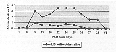
responders, 83% among slow responders, and 25% among non-responders. The patient we
present was a a rapid responder to glucocorticoid therapy (Fig. 1).
Finally, in the first prospective randomized study of ;lucocorticoid treatment in patients
in the fibroproliferative phase of ARDS, Meduri2' demonstrated improved gas exchange,
decreases in mufti-organ dysfunction scoring, increases in rapid extubation, and a
reduction in mortality in patients treated with methylprednisone compared with patients
receiving conventional treatment. Vasopressor dependence to compensate for septic shock is
another interesting point.21 Various studies have demonstrated increased mortality and
vasopressor dependence in septic patients with suprarenal insufficiency." Others have
reported patients without absolute suprarenal failure but with an insufficient ACTH
response whose prognosis and response to glucocorticoids were various." In the case
presented here, the patient did not present suprarenal failure, as demonstrated by the
basal cortisol level. He did not present relative suprarenal failure either, and the basal
cortisol level was therefore doubled in response to the ACTH challenge. However, the
administration of glucocorticoids led to rapid removal of epinephrine (Fig. 1). The
clinical use of glucocorticoids in septic shock has been practised for more than 30 years
but there is no conclusive evidence of their benefit as no well-designed studies have been
conducted."'Two recent meta-analyses demonstrated no benefit of the same, and noted
that such treatment could be deleterious. However, some studies have used glucocorticoid
treatment even in later stages of septic shock, with satisfactory results, noting an
improvement in haemodynamics and a decrease in vasopressor treatment." In Briegel's
study" patients who required catecholamiries for more than 48 h and received
glucocorticoids reversed shock within 7 days in 68% of cases, while patients in the
control group achieved this in only 21 % of cases. Mortality was 32% in the glucocorticoid
group and 63% in the control group. This study explicitly excluded patients with
suprarenal insufficiency and demonstrated that the response to glucocorticoids was similar
in both responders and non-responders to ACTH. It is possible to demonstrate that
glucocorticoids can be beneficial if used during the opportune period and that the
response to glucocorticoid administration is independent of the suprarenal reserve. To our
knowledge, this is the first report of successful treatment with glucocorticoids of a burn
patient presenting septic shock and ARDS.
|
Fig.1 -
Relationship between the Lung Injury Scale (LIS) and epinephrine requirements during
various stages of the clinical course. Adrenaline dose = mcg/kg/min. Day 9 = day
1 MV. Day 25 = day 1 methylprednisolone. Day 29 = e xtubation. Day 55
= Discharge. |
|
Conclusions
Both endogenous and exogenous
glucocorticoids protect the body from an exaggerated inflammatory response, an event that
can be more deleterious than the initial injury or trauma. It is clear that glucocorticoid
therapy cannot be administered haphazardly in ARDS or septic shock conditions. However,
glucocorticoids appear to be beneficial in the fibroproliferative stage of ARDS and septic
shock with controlled infection, when the inflammation response is out of control. This is
independent of the suprarenal reserve. In the patient we present the use of
glucocorticoids facilitated rapid weaning off mechanical ventilation and vasopressor
agents.
RESUME. Les
Auteurs presentent le cas d'un patient male atteint de brulures daps 23% de la surface
corporelle qui a developpe le syndrome de detresse respiratoire des adultes (SDRA) et le
choc septique pendant I'evolution. Wine si 1'infection etait maintenue sous controle,
1'insuffisance respiratoire et les besoins d'agents vasocompresseurs ne se sont pas
ameliores sans le traitement glucocorticoide. Ce type de traitement a permis d'interrompre
la ventilation mecanique et de suspendre les agents vasocompresseurs pendant la premiere
semaine du traitement. L'emploi des glucocorticoides dans les patients brines atteints
d'SDRA ou de choc septique est controverse, mais cc traitement pent se reveler efficace
dans la phase fibroproliferative de 1'SDRA et du choc septique si I'infection est sous
controle, comme dans le cas presente.
BIBLIOGRAPHY
American College of Chest
Physicians/Society of Critical Care 17.Medicine Consensus Committee: Definitions for
sepsis and organfailure and guidelines for the use of innovative therapies in sepsis.Crit.
Care Med., 20: 864-74, 1992.
Bone R., Fisher C.J., Clemmer R.P., Slotman
G.J., Metz C.A.:Early methylprednisolone treatment for septic syndrome and adult
18.respiratory distress syndrome. Chest, 92: 1032-6, 1987.
Hooper R.G., Kearl R.A.: Established ARDS
treated with a sustained course of adrenocortical steroids. Chest, 97: 138-143, 19.1990.
Soni A., Pepper G.M., Wyminski P.M.,
Ramirez E.N., Simon R.: Adrenal insufficiency occurring during septic shock: Incidence,
outcome, and relationship to peripheral cytokine levels. Am. J. Med., 98: 266-71, 1998.
Bernard G.R., Artigas A., Brigham K.L.,
Carlet J., Falke K.: The 21.American-European Consensus Conference on ARDS.
Definitions,mechanisms, relevant outcomes, and clinical trial co-ordination.Am. J. Resp.
Crit. Care Med., 149: 818-24, 1994.ti.
Bollaert P.E., Charpentier C., Levy B.,
Debouverie M., Audibert G., Larcan A.: Reversal of late septic shock with supraphysiologic
doses of hydrocortisone. Crit. Care Med., 26: 645-50, 1998.
Fiddian-Green R.G., Haglund U., Gutierrez
G., Shoemaker W.: Goals for the resuscitation of shock. Crit. Care Med., 21: S25-S31,
1993.
Luce J.M., Montgomery A.B., Marks J.D.,
Turner J., Metz C.A., Murray J.F.: Ineffectiveness of high-dose methylprednisolone in
preventing parenchymal lung injury and improving mortality in patients with septic shock.
Am. Rev. Respir. Dis., 138: 62-8, 1988.
Ashbaugh D.G., Maier R.V.: Idiopathic
pulmonary fibrosis in adult respiratory distress syndrome: Diagnosis and treatment. Arch.
Surg., 120: 530-5, 1985.
Briegel J., Forst H., Hellinger H., Haller
M.: Contribution of cortisol deficiency to septic shock. Lancet, 338: 507-8, 1991.
Chastre J., Fagon J.Y., Soler P., Bonnet
M., Domart Y.: Diagnosis of nosocomial pneumonia in intubated patients undergoing
ventilation: Comparison of the usefulness of bronchoalveolar lavage and the protected
specimen brush. Am. J. Med., 85: 499506, 1988.
Jones R.L., King E.G.: The effects of
methylprednisolone on oxygenation in experimental hypoxemic respiratory failure. J.
Trauma, 15: 297-303, 1975.
Meduri G.U., Chastre J.: The
standardization of bronchoscopic techniques for ventilator-associated pneumonia. Chest,
102 (suppl. 1): 557-64, 1992.
Meduri G.U., Headley S., Golden E., Carson
S.J., Umberger R.A., Kelso T., Tolley E.: Effect of prolonged methylprednisolone therapy
in unresolving acute respiratory distress syndrome. JAMA, 280: 159-65, 1998.
Montgomery A.B., Stager M.A., Carrico C.J.,
Hudson L.D.: Causes of mortality in patients with the adult respiratory distress syndrome.
Am. Rev. Respir. Dis., 132: 485-9, 1985.
Rottwell P.K., Udwadio Z.F., Lawler P.G.:
Cortisol response to corticotropin and survival in septic shock.. Lancet, 337: 582-3,
1991.
Artigas A., Bernard G.R., Carlet J.,
Dreyfuss D., Gattinoni L.: The American-European Consensus Conference on ARDS, part 2.
Ventilatory, pharmacologic, supportive therapy, study design strategies, and issues
related to recovery and remodeling. Am. J. Respir. Crit. Care Med., 157: 1332-47, 1998.
Lefering R., Neugebauer E.A.M.: Steroid
controversy in sepsis and septic shock. A meta-analysis. Crit. Care Med., 23: 1294-1303,
1995.
Hollingsed T.C., Saffle J.R., Barton R.G.,
Craft W.B., Morris S.E.: Etiology and consequences of respiratory failure in thermally
injured patients. Am. J. Sung., 166: 592-7, 1993.
Cronin L., Cook D.J., Carlet J.:
Corticosteroid for sepsis: A critical appraisal and meta-analysis of the literature. Crit.
Care Med., 23: 1430-9, 1995.
Meduri G.U., Chinn A.J., Leeper K.V.,
Wunderink R.G., Tolley E.. Corticosteroid rescue treatment of progressive
fibroproliferation in late ARDS. Pattern of response and predictors of outcome. Chest,
105: 1516-27, 1994.
Peck M.D., Weber J., McManus A.:
Surveillance of burn wound infections: A proposal for definitions. J. Burn Care Rehabil.,
19:386-9, 1998.
Hakkinen P.J., Schomoyer R.L., Wtischi
H.P.: Potentiation of butylated-hydroxytoluene-induced acute lung damage by oxygeneffects
of prednisolone and indomethacin. Am. Rev. Resp. Dis., 128: 648-5, 1983.
Sprung C.L., Caralis P.V., Marcial E.H.:
The effects of high-dose corticosteroids in patients with septic shock. N. Engl. J. Med.,
31: 1137-43, 1984.
Schuster D.P., Kollef M.H.: Acute
respiratory distress syndrome.Disease a Month., 5: 270-325, 1996.
Veterans Administrations Systemic Sepsis
Co-operative Study Group. Effect of high-dose glucocorticoid therapy on mortality in
patients with clinical signs of systemic sepsis. N. Engl. J. Med., 317: 659-65, 1987.
Bernard G.R., Luce J.M., Sprung C.L.:
High-dose corticosteroids in patients with the acute respiratory distress syndrome. N.
Engl. J. Med., 317: 1565-70, 1987.
Meduri G.U., Belenchia J.M., Estes R.J.,
Wanderink R.G., Torky M.E., Leeper K.V.: Fibroproliferative phase of ARDS. Clinical
findings and effects of corticosteroids. Chest, 100: 943-52, 1991.
Murray J.F., Matthay M.A., Luce J.M.: An
expanded definition of the adult respiratory distress syndrome. Am. Rev. Respir. Dis., 38:
720-3, 1988.
| This paper was received on 3
October 2000 Address
correspondence to:
Dr C. Ramos CCEPAQ, Benaim Foundation,
Aleman Hospital, Buenos Aires, Argentina. |
|