Annals of Burns and Fire Disasters - vol. XIV - n. 1 - March 2001 MARJOLIN'S ULCER OF THE SCALP: CASE REPORT AND LITERATURE REVIEW Malheiro E., Pinto A., Choupina M., Barroso L., Reis J., Amarante J. Servigo Cirurgia Plastica, Hosp. S. Joao,
Porto, Portugal SUMMARY. This paper presents the case of a 32-year-old woman with a large, infected, and ulcerated squamous cell carcinoma in the parieto-occipital region. A review of the literature regarding patients with burn scar carcinoma is also presented. History The earliest identification of the malignant degeneration of burn scars is attributed to Celsus, early in the first century.' Subsequent reports of carcinoma arising in post-traumatic scars include the classic description by a French physician, Jean-Nicholas Marjolin, in 1828.2 Da Costa in 1903 was the first author to use the eponym "Nfarjolin's ulcer" to describe the skin cancers occurring in burn scars.' Although most commonly seen in burn patients, Mau-john's ulcers are also reported in previously traumatized and scarred tissue of various aetiology, such as osteomyelitis,' frostbite,' venous stasis ulcers,' skin graft donor sites,' chronic pressure ulcers,' poorly fabricated prostheses,' gunshot wounds, puncture wounds, dog bites," and blunt trauma."
Skin subjected to repeated thermal insults has long been recognized to have an increased incidence of carcinoma. There are two populations where a similar malignant change is common. The Indian Kangri ulcer, prevalent among the poorer classes of Kashmir, is a consequence of wearing warm embers. The Kairo burn cancer seen in Japan results from burn scars due to wearing a small tin oven underneath the clothing." However, there seems to be no predilection for particular races.' Malignant changes in burn scars may occur at any age. Seen predominantly in adults, burn scar carcinoma occurs at an average age of 53-59 yr." As a general rule the typical time lag between the burn and development of cancer is between 25 and 40 yr."'," Another interesting observation is that the latent period is inversely proportional to the patient's age at the time of the burn. The younger the patient at the time of injury, the longer the interval for malignant change, while the older the patient at the time of burning, the longer the lag period. '5 Although burn injuries are commoner in females, the majority of studies refer that males present an incidence three times greater than females."-'8 The reason for this phenomenon is unknown. Most Marjolin ulcers occur on the extremities. The anatomical locations reported by Arons," Lawrence," and Novick" show that average distribution of Marjolin ulcers is 40% in the lower extremity, 30% in the head and face, 20% in the upper extremity, and 10% in the trunk area. Several researchers have pointed out the propensity of burn scar cancer to arise in flexion creases.','' This distribution is in marked contrast to the distribution of spontaneous epidermoid carcinomas, which are located on the face and neck in more than 90% of cases. `20 In a clinical study by Hahn et a1." it was found that burns were the commonest antecedent trauma, the second commonest cause being chronic osteomyelitis. It has also been estimated that Marjolin ulcers occur in 1.5% of all osteomyelitis patients, and in 2% of all burn Victims. 1,22 One study reports an incidence of 1:1000 occurrences in patients with chronic venous stasis ulcers," Squamous cell carcinomas are by far the commonest type of cancer arising from Marjolin ulcers, with basal cell carcinomas coming second. 1,11,21 Other neoplasms have been reported arising from chronic ulcers; these include melanoma ,z5 osteogenic sarcoma," fibrosarcoma,' liposarcoma, carcinosarcoma, and carcinoma in situ.', Pathophysiology Although Arons reported three exceptions, it is generally accepted that burn scar cancer very seldom arises when burn wounds have been primarily grafted." The exact mechanism of the malignant conversion occurring in chronic ulcers is still not known. As with all unanswered questions, many theories have been proposed. Among the theories advanced for the pathogenesis of Marjolin's ulcer are Virchow's theory" of chronic irritation, Ribbet's theory" of misplaced epithelial cell groups, and those of Treves and Pack, who suggest that as a result of the constant breakdown of the ulcer a nutritional deficiency develops, owing to the release of toxins by autolysis and heterolysis of the burn scar. This yields an epithelium that is unable to withstand the carcinogens produced by the skin because of excessive heat and radiation." Another explanation is that owing to the malnourished tissue present, chemical damage is again poorly tolerated and mutates DNA genes, which never undergo repair." Castillo and Goldsmith believed that owing to the obliteration of lymphatics in scar tissue, a poor antigen-antibody response was seen in both local and regional lymph nodes." Futrell and Myers later added to this theory, suggesting that this allowed the tumour to "escape" recognition by the normal host. The tumour was thus enabled to reach a critical size before detection took place." Studies have also shown patients with Marjolin ulcers have a decreased T-cell count, suggesting that immunosuppression is a contributory factor.' Whatever the exact mechanism, most researchers agree that a "cancerous environment" is formed because of the lack of blood supply and immunity in the scar tissue. The dry, thin, delicate epithelium covering the scar is easily destroyed by the trauma to which it is frequently subjected. The epithelium may easily be abraded by slight injuries, movement over a joint or across a flexion crease, or prolonged itching. Each successive abrasion, ulceration, or fissure heals with increased difficulty and the regenerated epithelium is progressively inferior. Ultimately, persistent stimulation to the marginal epithelium for regeneration and repair, with constant frustration, may lead to a loss of tissue restraint and eventually to neoplastic changes. Clinical diagnosis Marjolin ulcers generally occur in regions of previous deep burn that healed slowly without skin grafting. It is widely believed that the signs and symptoms associated with a cancerous ulcer are commonly mistaken for an infection rather than a malignancy." Burn scar carcinoma presents as an indolent, flat, chronic ulcer enlarging in circumference and depth, with indurated and elevated margins and a granular base. The ulcer is foul-smelling and painful, with an alteration in the quantity of exsudate and a bloody drainage content'"" Additionally, radiographic films may show bone destruction.' Biopsy remains the gold standard for the diagnosis of malignant change. The procedure should be performed at multiple sites including central areas as well as margins. If the biopsy report suggests atypical pseudoepitheliomatous hyperplasia, we should be on our guard, as this has been linked to a possible malignant change, and it is often difficult to distinguish from squamous cell carcinoma.'.." It has been suggested that if biopsy reports reveal atypical pseudoepitheliomatous hyperplasia, repeated biopsies should be performed at three-month intervals until complete healing has occurred. Lastly, regional lymph nodes should be palpated for possible enlargement due to metastasis. Several studies have shown that approximately 30% of Marjolin ulcers have associated lymphadenopathy Treatment and prognosis There is a measure of agreement regarding several basic principles in the treatment of Marjolin ulcers. First of all, the best treatment for burn scar cancer is preventive medicine. Once full-thickness bums occur they should be skin-grafted at the optimal moment since, generally speaking, malignant degeneration rarely develops in a primarily grafted burn. As in all wounds, infection should be addressed early. If drainage is present it should be handled appropriately. In most cases, chronic ulcers should be excised, even if they are benign, and appropriate skin grafts or flaps should be used. It has been suggested that the excisional boundaries should include 3-4 cm of surrounding normal skin tissue including muscle fascia." Kipikasa and Guzanin expressed the opinion that the excision should always be performed by cautery." This was due to their belief that surgery was the critical breaking point at which tumour cells could be washed into the bloodstream and lymphatics, leading to metastasis. This theory helps to explain the unusually high occurrence of metastasis after surgery. Amputation is now usually reserved for deep ulcerated lesions which extend into joint cavities and bone." However, some believe it should be standard treatment in all cases." Controversy continues to exist with regard to lymph node dissection. Many researchers have advocated prophylactic dissections."` However, the majority of studies recommend that lymph node dissection should be performed only when the nodes are clinically palpable.' Whatever the treatment, long-term follow-up is recommended in all cases. One study cited a 98% metastatic recurrence rate within 3 years of local excision." Most studies have indicated a recurrence rate ranging between 20 and 50%, with regional lymph nodes being the commonest sites affected, followed by the brain, liver, kidney, and lungs."" If the regional lymph nodes become involved, the 3-year survival rate decreases to 35%." Novick et al. found a 54% metastatic occurrence from Marjolin ulcers in the lower extremity." This rate was more than two times as high as the metastatic rate in any other site. Barr and Menard reported that patients with lesions in the lower extremities presented a higher rnetastasis rate and a lower 5-year survival rate than patients with lesions in any other primary site." It has been reported that patients who survive three years without metastasis have an excellent prognosis Case report We present the clinical case of a 32-year-old Caucasian vioman with
a large, infected, and ulcerated squamous cell carcinoma mass in the parieto-occipital
region who was referred to our Plastic and Reconstructive Centre. At the time of
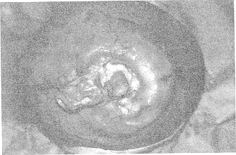
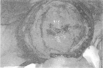
presentation, the patient presented a yellowish, keratotic, vegetative mass of hard
consistency, measuring 4.5 x 4 cm. Nearby there was a deep, whitish ulcer (4 x 4 cm)
invading the cranium and exposing the dura mater. Its borders were indurated and elevated.
The ulcer had a foul odour and a sticky yellow exsudate (Figs. 1, 2). The chest x-ray
appeared normal.The tumour was shown by CT to have invaded the cranium (Fig. 3). RMN did
not reveal cervical lymphadenopathy. Other laboratory studies were found to be within
normal limits. An arteriography did not show invasion of the superior sagittal sinus.
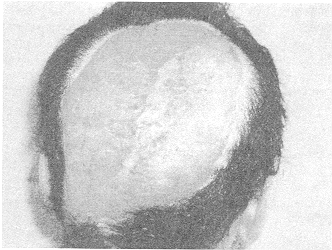
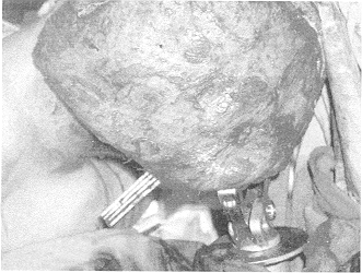
Surgical resection was performed with wide margins, including the full thickness of the
skull (Fig. 4). A free latissimus dorsi muscle flap measuring 19 x 29 cm was used to cover
the defect (Fig. 5). No bone reconstruction was performed. One week later the flap was
split-thickness grafted. The specimen confirmed the diagnosis of squamous cell carcinoma.
The margins were free of neoplasm. The postoperative course was uneventful and all the
surgical sites healed (Fig. 6). The patient was disease-free at six months' follow-up.
Conclusion This paper was written in order to highlight the existence of a condition that is commoner than we may think and needs to be correctly diagnosed and treated by plastic and reconstructive surgeons. Marjolin's ulcer is not a rare phenomenon - physicians should be aware of its prevalence after burn trauma and refer cases to burns centres earlier than they usually do. The clinical signs and symptoms are often mistaken for infection, and misdiagnosis may occur. In a condition that can rapidly become devastating for the patient, clinicians must today be aware of a benign ulcer's potentiality to turn malignant. Also, prompt aggressive treatment must be initiated before metastasis occurs.
RESUME. Les Auteurs considerent le cas dune femme de 32 ans atteinte d'un epithelioma spinocellulaire etendu, infecte et ulcere daps la region parieto-occipitale. Its presentent en outre la litterature sur les patients atteints de carcinome des cicatrices de brulure. BIBLIOGRAPHY
|