Annals qf ihe MBC - vol. 3 - n' 4 -
December 1990
HIGH-TENSION
ELECTRICAL BURNS. PRIMARY TREATMENT OF SEVENTY PATIENTS
Escudero-Nafs F.J., Leiva-Oiiva R.M.,
Collado-Aromir F, Rabanal-Suirez F, De Molina-NOfiez 1M.
Department of Plastic Surgery and Burns, Valle de
Hebron. General Hospital, Barcelona, Spain
SUMMARY.
Seventy patients with high-tension bums, admitted to the <<Valle de Hebron>>
General Hospital Burn Unit, during a period of 9 years (1980-1988), were evaluated
retrospectively. Only victims with documented passage of high-tension electrical current (
> 1,000 volts) through the body were reviewed. Patients with only flash or flame bums
from electrical accidents were excluded. There were 69 males and I female, with an average
age of 31.3 years. Thirty-nine accidents (55.7%) occurred at work, and 31 (44.3%) occurred
in leisure time. All the patients, with one exception, were admitted within 5 hours of
injury. The extremities were the most frequent site of injury. In 54 cases (77%), two or
more extremities were burned. Thirty patients (43%) sustained additional flash and/or
flame bums. The mean total body surface area of the burns was 12%, with a mean
full-thickness bum area of 7%.
Twelve patients (17%) required
cardiopulmonary resuscitation at the scene of the accident or following admission. Fluid
resuscitation was carried out using Ringer's lactate at a vigorous rate. Acidotic
patients, or with marked myoglobinuria, were additionally treated with intravenous sodium
bicarbonate, mannitol or both. A total of 179 operations were performed, for exploration,
debridement and closure of the injuries. Thirty-six cases'(51.4%) underwent a total of 56
amputations; 14 patients suffered multiple amputations.
Acute renal failure developed in two cases (2.8%), and both died. No cases of clostridial
infection occurred. Sixty-seven patients (95.7%) were discharged, with an average stay of
50 days. Three deaths occurred among the reviewed cases (4.3%). We attribute this low
mortality to early transfer of patients to our Burn Unit, aggressive fluid resuscitation,
and early aggressive debridement.
Introduction
Electricity in its unprotected, naked
form may produce both death and devastating injuries (1). Electrical burns account for 3
to 17 per cent of all admissions to Burn Centres (2, 3).
Although there are several factors that decide the severity of the traumatism, the
principal determinant of tissue damage is the voltage of the current (4). Traditionally,
injuries caused by exposure to 1,000 volts or greater are considered high-tension
electrical bums (1, 2). Luce and Gottlieb (5) have divided these injuries into flash and
"true" high-tension electrical burns. The former are caused by a flame of very
high temperature and short duration, due to the conversion of electrical energy to heat
outside the body (6). These burns are typically mixed full- and part ial-thickness burns,
without characteristic entry and exit wounds. "True" electrical injuries are
caused by the passage of an electrical current through the body, between entrance and exit
points, with significant destruction in these areas. Both flash and "true"
electrical burns may occur in the same patient. Secondary burns can be inflicted by
ignition of clothing or environmental objects.
High-tension electricity may produce massive tissue loss. In addition to cutaneous
injuries, there is severe damage to underlying muscles, nerves, blood vessels and bones,
which frequently results in amputations, renal failure and other complications. Every
organ system can be injured by the passage of current. Vigorous resuscitation, with early
wound exploration and debridement, may prevent some of the devastating disabilities and
systemic complications of these burns. In this paper, we present a review of the treatment
of high-tension electrical burns in our Department.
Material and methods
Seventy patients with high-tension
electrical burns, admitted during a period of 9 years (1980-1988), were evaluated
retrospectively. Only victims with documented passage of high-tension electrical current
(>1.000 volts) through the body were included in this review. Patients with only flash
or flame burns from electrical accidents were excluded.
There were 69 males and 1 female, with an average age of 3 1.3 years (range: 8 to 75).
Thirty-nine patients (55.7%) were injured at work; 23 of these cases were electricians and
6 were construction workers. Thirty-one accidents (44.3%) occurred in leisure time. The
majority of accidents were caused by grasping or touching high-voltage lines, or happened
in transformers or electrical substations. Lightning produced injuries to one patient.
The mean voltage involved was 15,000 volts (range: 8,000 to 25,000); the voltage of the
lightning was unknown.
All the patients, with one exception, were admitted within 5 hours of injury.
All the patients sustained entrance and exit wounds (Figs. I and 2). The extremities were
the most frequent site of injury (Tab. l). In 54 cases (77%), two or more extremities were
burned (Tab. 2). In addition to the "true" electrical injuries, 30 patients
(43%) sustained flash and/or flame bums. The mean total body surface area of the bums was
12% (range: 2 to 45), with a mean full-thickness bum area of 7% (range: I to 40). Five
cases had fractures with soft tissue wounds.
Treatment. Twelve patients (17%) required cardiopulmonary resuscitation at the
scene of the accident or following admission; all these victims survived.
Electrocardiographic monitoring was performed if clinical evaluation so indicated.
Fluid resuscitation was carried out using Ringer's lactate. The rate of fluid
adminstration was adjusted to keep a urine output of I to 1.5 ml/kg body weight per hour.
Acidotic patients, or with marked myoglobinuria (41 cases, 58,5%), were additionally
treated with intravenous sodium bicarbonate, mannitol, or both. Ringer's lactate was
continued at a vigorous rate to grossly clear the pigments.
After initial stabilization, patients with signs of increasing compartment pressures or
myonecrosis were taken to the operating room (usually within 4 hours after admission), for
fasciotomies and tissue exploration. All potentially involved muscle compartments were
explored, including deep muscle groups adjacent to bone (Fig. 3). Obviously nonviable
tissues were debrided initially; amputations were performed only in clearly necrotic
extremities. Amputation sites were left open unless obviously viable tissue was present at
the level of amputation. Following initial exploration and debridement, exposed tissues
were covered with skin homografts, topical agents, or dressings soaked in physiological
serum. Silver sulphadiazine was used on the associated flash and flame bums. Wounds were
explored and debrided at 48 to 72 hour intervals, until all nonviable tissues were removed
or amputation was necessary. Wounds were then closed with skin autografts, flaps or both.
Associated full-thickness flash and flame bums were treated through early excision and
grafting.
Rehabilitation was started during the hospital stay and continued after discharge.
Results
Surgical procedures. The
patients reviewed required a total or 179 operations, ranging from one to six surgical
procedures per patient (mean number 2.5), for exploration, debridement and closure of the
injuries. Thirty-six cases (51.4%) underwent a total of 56 amputations; removal of
multiple fingers or toes form the same extremity counted as one amputation. Almost two
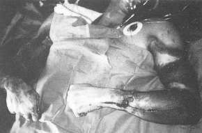
thirds of the amputations involved the upper limbs. Fourteen patients had multiple
amputations (Figs. 4 and 5). One bilateral shoulder disarticulation and two unilateral
shoulder disarticulations were performed (Tab. 3).
 |
 |
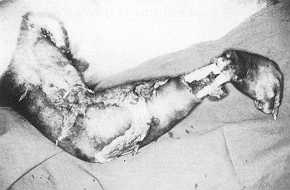
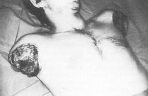
| Fig. 1 A
25-year-old man with entrance injuries of a high-tension electrical bum. The current
vaporized the soft tissues in the wrist and distal forearm. Shoulder disarticulation was
performed. |
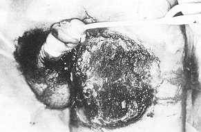
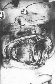
Fig.
2 Exit wounds of a high-tension electrical bum, involving the left thigh, penis
and scrotum. |
|
A right fourth rib excision was necessary
in one case with electrical burns on the chest wall. One patient sustained a
full-thickness loss of the abdominal wall, with prolapse of perforated intestinal loops
through the wound; in addition to debridement of the burns, the necrotic intestine was
resected, and intestinal anastomosis was performed (Fig. 6).
Surgical procedures used for closure of the electrical injuries are shown in Tab. 4.
| Location |
Entrance
site
(N' of cases) |
Exit site
(N' of cases) |
| Right upper extremity |
46 |
3 |
| Left upper extremity |
37 |
5 |
| Right lower extremity |
5 |
33 |
| Left lower extremity |
3 |
37 |
| Head and neck |
3 |
7 |
| Trunk |
7 |
4 |
| Penis and scrotum |
0 |
3 |
|
Table 1 Entrance
and exit sites of electrical burns |
|
Complications and mortality. Two
patients developed acute pulmonary oedema from fluid overload. One of these sustained an
acute gastrointestinal haemorrhage, as a result of stress ulcers. Acute renal failure
developed in only two patients (2.8%), both of whom died. No instances of infection with
clostridial organisms occurred.
There were three deaths (4,3%). The mean age of the deceased patients was 40 years, and
the mean total body surface area of the burns was 20%. One victim suffered a deep
electrical bum on his scalp and skull (Fig. 7). He died nine days after injury with severe
cerebral oedema and acute respiratory insufficiency.
 |
 |
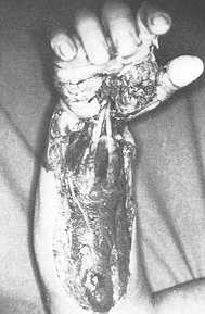
| Fig. 3 High-tension
electrical bum of the right upper extremity: Decompressive fasciotorny of forearm, with
extension of surgical incision through the carpal tunnel space of the hand. |
Fig.
4 An 18-year-old boy with high-tension electrical burns involving upper
extremities. |
 |
 |
| Fig.
5 Patient of figure 4. Bilateral amputation of upper extremities following
high-tension electrical bums. |
Fig. 6 A
45-year-old man with extensive destruction of the abdominal wall by high-tension
electrical current. The patient died three days after injury. |
|
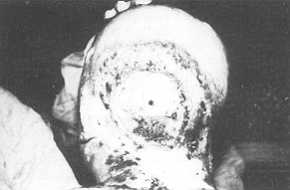 |
Fig. 7 High-tension
electrical bum of the scalp, skull and neck. The patient died nine days after injury. |
|
The patient with destruction of the
abdominal wall and intestinal necrosis had severe electrical bums on his left leg. An
above-knee amputation was performed. He died three days after injury, as a result of
peritonitis, generalized sepsis and acute renal failure. A 50-year-old man had an
associated ,skull fracture. Following exploration and debridement of the bums, the patient
died ten days after injury. He developed multiple organ failure, including acute renal
failure.
Length of hospitalization. Sixty-seven patients (95.7%) were discharged from the
13th to the 112th days following admission (average stay, 50 days).
Discussion
Patterns of high-tension injuries
include instant death, massive tissue loss, secondary ignition burns, and associated
traumatisms. The victims are almost exclusively young men, with an average age of 30
years. Approximately one third of these injuries occur in electrical workers (1, 4).
Resuscitation following electrical accidents must be directed immediately to the
cardiorespiratory systems, because respiratory paralysis and ventricular fibrillation are
the principal causes of death (4). Because the victims tend to be young and in good
health, prolonged efforts at cardiopulmonary resuscitation are warranted (7).
One of the major complications of high-tension electrical bums is acute renal failure.
Circulating muscles and red blood cells may lead to acute tubular nephropathy, in the
presence of low renal blood flow and low urine volumes. Prompt fluid resuscitation is the
key to the prevention of this complication. myoglobin and haemoglobin from damaged Because
of the iceberg effect of the cutaneous injury, coupled with the extensive destruction of
all underlying structures, the fluid requirements are much greater than in a comparable
thermal bum (1, 4, 8). Large infusions of Ringer's lactate should be administered, at a
rate that maintains a urine output of approximately I to 1.5 ml/kg body weight per hour.
The use of mannitol to increase renal perfusion and sodium bicarbonate to achieve urine
alkalinity has been an integral part of the resuscitation programme for electrical bums
(1, 2, 3, 9, 10).
Sometimes it is impossibile to resuscitate a patient with a large amount of dead muscle,
and an emergency operation to amputate the injured extremity or remove the destroyed
tissues may be necessary (11).
The incidence of acute renal failure in most series has been reported to be 1.5 to 7.5 (2,
12). Two cases (2.8%) of this complication occurred in our series.
After the cardiopulmonary and renal situations have stabilized, attention must be directed
to the injured tissues. We agree with Holliman et al. (13) that early and repeated direct
inspection of all damaged muscle groups seems to be the most reliable method of assessing
vaibility. Parshley et al. (14) have emphasized the importance of immediate decompression
of tight muscle compartments, by early and radical fasciotomy, to prevent further damage
to the tissues, and simultaneous and often radical removal of obviously nonviable muscles,
including immediate amputation of extremities that are clearly not salvageable. Care must
be taken to investigate not only the superficial muscle layers, but also the deep muscle
adjacent to the bone (4). Muscles with questionable viability should be left for
re-exploration and re-evaluation at 48 to 72 hour intervals (13, 14). Nerves and tendons
should be preserved even if they appear devitalized (2). Luce et al. (15) have observed
regeneration and return of function in apparently nonviable nerves. Preservation of flexor
tendons can facilitate later tendon transfer during the process of reconstruction.
Repeated debridements are usually necessary in order to excise all devitalized tissues.
Definitive coverage is provided when no further nonvital tissue surgeons (16, 17) believe
that tissue can be salvaged by early coverage with a flap that brings new blood supply,
but others feel that flaps covering nonviable tissue may become necrotic, and serve as a
source of severe clostridial infection (4). Coverage may be achieved by means of
autogenous split-thickness skin grafts, but they do not provide the stability often
required (2). If a substantial number of exposed muscles, tendons, and nerves are deemed
viable at the initial or subsequent procedures, coverage within the first few days by a
pedicle or free flap may preserve essential function in a way that no other method can
achieve (1, 15, 18).
Wang et al. (19) have obtained satisfactory results in the prevention of limb necrosis
using early vein grafts for re-establishing blood circulation in wrists showing impaired
blood flow, as a result of electrical injury.
Amputation should be carried out as soon as it becomes clearly necessary, both to lessen
the risk of later invasive infection, and to lessen the load of myoglobin and tissue
toxins that could be absorbed by the bloodstream (14). Remensnyder (1) has summarized some
of the reported experience with major amputations in high-tension electrical injury. Of
the 598 patients reported, 37% underwent one or more major amputations. A total of 56
amputations were performed in 36 cases (51.4%) of our review; 36 were major amputations.
The overall mortality rate from electrical bums has been reported to be between 3 and 14%
(2, 6, 9). Our mortality rate was 4.3%. We attribute this low incidence to early transfer
of patients to our Bum Unit, aggressive fluid resuscitation and early aggressive
debridement.
RÉSUMÉ. Les
Auteurs ont exécuté une analyse rétrospective de 70 patients (69 mâles, 1 féminin,
âge moyen 31,3 ans) qui ont subi des brûlures électriques de haute tension (--1000 V)
hospitalisés pendant la période 1980-89 chez l'Unité des Brûlures de l'Hôpital
Général "Valle de Hebron". Pour ce qui concerne le lieu des accidents, 39
(55,7%) se sont produits à la maison et 31 (44,3%) au travail. Tous les patients, sauf
un, ont été hospitalisés dans les premières 5 heures après l'accident. Les
extrémités ont été la partie du corps plus fréquemment atteinte (deux ou plus de deux
extrémités en 54 cas (77%". Les Auteurs décrivent les thérapies employées et les
interventions chirurgicales effectuées. Ils attribuent le taux de mortalité très
limité (seulement trois patients sont décédés, c'est-à-dire 4,3% des cas) à
l'hospitalisation précoce chez leur Unité des Brûlures, à la réanimation agressive
avec l'emploi des liquides, et au débridement précoce et agressif
BIBLIOGRAPHY
- Remensnyder J.P.: Acute electrical injuries. In
J.A.J. Martyn (Ed.), "Acute Management of the Burned Patient", 66-86, W.B.
Saunders Company, Philadelphia, 1990.
- Luce E.A.: Electrical injuries. In: J.G. McCarthy
(Ed.), "Plastic Surgery", 814-830, W.B. Saunders Company, Philadelphia, 1990.
- Haberal M., Kaynaroglu V., Oner Z., Gulay H.,
Bayraktar U., Bilgin N.: Epidemiology of electrical bums in our centre.Annals of the MBC,
2: 14-16, 1989.
- Bingham H.: Electrical bums. Clin. Past. Surg., 13:
75-85, 1986.
- Luce E.A., Gottlieb S.E.: "True"
high-tension electric injuries.Ann. Plast. Surg., 12: 321-326, 1984.
- Gordon M.W.G., Reid W.H., Awwaad A.M.: Electrical
bums. Incidence and prognosis in Western Scotland. Bums, 12: 254-259, 1986.
- Salisbury R.E., Dingeldein G.P. Jr.: Specific bum
injuries. In: Salisbury R.E., Newman N.M., Dingeldein G.P. Jr. (Eds.), "Manual of Bum
Therapeutics. An Interdisciplinary Approach", 81-86, Little, Brown and Company,
Boston, 1983.
- Rouse R.G., Dimick A.R.: The treatment of electrical
injury compared to bum injury: a review of pathophysiology and comparison of patient
management protocols. J. Trauma, 18: 43-47, 1978.
- Wilkinson C., Wood M.: High voltage electric injury.
Am. J. Surg., 136: 693-696, 1978.
- Arturson G., Hedlund A.: Primary treatment of 50
patients with high-tension electrical injuries. I. Fluid resuscitation. Scand. J. Plast.
Reconstr. Surg., 18: 111-118, 1984.
- Artz C.P.: Electrical injury. In: Artz C.P.,
Moncrief J.A., Pruitt B.A. Jr., "Bums: A Team Approach", 351-362, W.B. Saunders
Company, Philadelphia, 1979.
- Hanumadas M.L, Voora S.B., Kagan R.J., Matsuda T.:
Acute electrical bums: a 10-year clinical experience. Bums, 12: 427-431, 1986.
- Holliman C.J., Saffie J.R., Kravitz M., Warden G.D.:
Early surgical decompression in the management of electrical injuries. Am. J. Surg., 144:
733-739, 1982.
- Parshley P.F., Kilgore J., Pulitc, J.F., Smiley
P.W., Miller S.H.: Aggressive approach to the extremity damaged by electric current. Am.
J. Surg., 150: 78-82, 1985.
- Luce E.A., Dowden W.L., Su C.T., Hoopes J.E.:
High-tension electrical injury of the upper extremity: Surg. Gynecol. Obstet., 147: 38-42,
1978.
- Jenkins A.M., Pegg S.P.: Island flaps in the primary
reconstruction of electrical bums. Bums, 13: 236-240, 1987.
- Govila A.: Early excision and primary resurfacing of
wounds following high voltage electrical burns. Eur. J. Plast. Surg., 12: 147-154, 1989.
- Silverberg B., Banis J.C. Jr., Verdi G.D., Acland
R.D.: Microvascular reconstruction after electrical and deep thermal injury. J. Trauma,
26: 128-134, 1986.
- Wang XW., Roberts B.B., Zapata R.L., Robinson W.A.,
Waymack J.P., Law E.J., MacMillan B.G., Davies JW.L.: Early vascular grafting to prevent
upper extremity necrosis after electrical bums. Commentary on indications for surgery.
Bums, 11: 359-366, 1985.
|