Annals of 1he MBC - vol. 5 - n' 3 -
September 1992
A
DIFFERENTIAL MODEL OF WOUND EPITHELIALIZATION FOR FULL-THICKNESS SKIN DEFECTS
Manios A., Tzortzak¡s H., Minogiannis N., Tsiftsis D.
Clinical Information Sciences Laboratory, Surgical
Department, Crete University, Greece
SUMMARY.
A differential model of wound epithelialization is presented which describes
concurrently the two processes of epithelial cell migration and epithelial cell mitosis.
The model is a non-linear differential equation which describes the wound
epithelialization process on the x-y plane. The equation has two components, a diffusion
component and a population dynamics component. The results of the computer simulation were
very realistic and will help the plastic surgeon to develop strategies for wound coverage
via the use of a personal computer.
In this work we present a mathematical
model of wound epithelialization. Our model is a differential equation with partial
derivatives which describes the behaviour of the most important variables involved in the
process of wound epithelialization. It is clear that the model itself is not the reality,
but it can simulate the main characteristics of the process under consideration (1).
Wound epithelialization is an extremely complex process that is difficult to simulate
precisely by any differential model because the phenomenon presents unclear points.
Nevertheless, it is possible to isolate the basic components of this process in order to
make a satisfactory model for study. This will help the investigator to understand the
process better and make conclusions about the efficiency of various techniques used to
manage skin defects. The classic process of building an experimental animal model of wound
epithelia] ization is not a rational approach because there are significant differences
between man and animals in this phenomenon. Mathematical modelling in this case is an
alternative method of study (1). Using some techniques of mathematical analysis it is
relatively easy to simulate the two main processes of wound epithelialization. As
characteristically described in Peacock's classic book "Wound Repair" (2), these
two processes are epithelial cell migration and epithelial cell mitosis. It is known that
after trauma there is a migration of cells from the basic layer of the epidermis towards
the defect. Later there is also some mitotic activity of these cells. After detailed
analysis we found that both processes can be described by a differential equation with
partial derivatives. This equation has two components, a diffusion part and a population
dynamics part, and the form:
where: u = cell concentration in an elementary area AxAy,
t time, k,c,u,, = constants
Eden in 1961 introduced a simple
mathematical model to describe tumour growth (3). The diffusion equation is well known to
engineers, physicists and biochemists. It describes heat transfer and the phenomenon of
diffusion. Cell mitosis can be described by the logistic equation. This equation is the
population dynamics part of our model. If we divide the skin defect into the elementary
surfaces Axay it is possible to compute - on the basis of the equation of our model - the
cell concentration in each area. This can be done if we know the geometry and the boundary
conditions of the defect. If the techniques of coverage of the defect alter the internal
and external boundaries in known manner it is easy to compute the healed area in each
period of time. The analytical solution of our equation is difficult, if not impossible.
We must therefore solve this equation numerically (4) with a computer. This can be done
using the finite difference approximation. This converts our differential equation into a
difference equation. Another method of solution is to convert our continuous model into a
discrete dynamic system (5).
The point which is interesting to the plastic surgeon in our model is that it permits the
comparison of the various methods used to cover skin defects. In our computer study we
used a rectangular skin defect of 100 area units using 36 units of skin. We used the same
quantity of skin as one segment, as 9 segments, as 36 segments and as a meshed skin graft
(1 x 2.7). We found that the most efficient method of coverage was 36 segments. This means
that the division into 36 segments covered more defect area in the same time (Fig. 1).
We selected the coefficients of diffusion and cell multiplication arbitrarily. It is not
the absolute value Wound[ epithelization and skin grafting of diffusion coefficient or the
absolute value of cell multiplication coefficient which affects the comparison of the
various techniques. It is the ratio between them. In order to see the difference between
them we altered the ratio from no mitosis to high mitotic activity in comparison to cell
diffusion. In all cases the results were the same as the multiple division model, becoming
stronger with high mitotic activity.
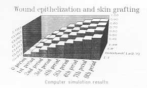 |
Fig. I.
Comparison between the various methods of wound coverage from computer simulation
results. |
|
These results would indicate that by
dividing a small skin graft into an enormous number of segments one would be able to cover
a very extensive defect. This would be reasonable if the dividing organ did not destroy
cells from the graft. In multiple divisions the number of destroyed cells will therefore
be unacceptable. The optimal number of divisions has to be defined by clinical
investigation.
One can easily understand the results of computer simulation by a simple fact. If we
divide a piece of skin and distribute the skin segments homogeneously in the defect area,
the cells from these grafts have to cover a smaller distance compared to those of one
piece placed in the centre of the defect. Morover the diffusion surface is higher with
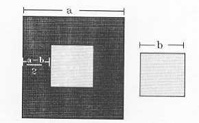
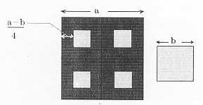
multiple segments than with just one. Our model verifies these facts mathematically (Fig.
2, 3).
We must emphasize that this kind of investigation depends on the hypothesis that a
biological system can be simulated by a computer. Computer simulation of biological
processes is a field in which many advances have been made in recent years. There are
extensive references to tumour models, population models and ecological systems in the
field of computer simulation. In 1987, Stevanovska et al. from Lubliana (6) described a
wound-healing model based on experimental data. Their model is an exponential equation
which describes the surface of an ulcer with time. This is similar to our model when we
run it for a defect which heals spontaneously.
Nowadays, computer simulation is an acceptable method of investigation in many centres.
Computer simulation can save money and time by obviating the use of experimental animals
without significantly affecting the accuracy of investigation.
 |
 |
| Fig. 2 If
the skin graft is used as one segment the diffusion line will be 4b. |
Fig. 3
If the skin graft is used as four segments the diffusion line will be 8b. |
|
RÉSUMÉ. Les auteurs présentent un modMe
différentiel de l'épithélialisation de la lésion qui décrit coneurremment les deux
procédés de la migration des cellules épithéliales et la tuitose des cellules
épithéliales. Le modéle est une équation différentielle non linéaire qui décrit le
procédé de Pépithélialisation de la lésion sur le plan x-y. L'équation á deux
composants, un composant de diffusion et un composant de dynamique de la population. Les
résultats de la simulation sur ordinateur se sont montrés trés réalistes et i1s
aideront le plasticien á déveloper des stratégies pour couvrir les lésions avec
l'emploi d'un ordinateur.
BIBLIOGRAPHY
- Andrews J.G., McLone R.R.: "Mathematical
Modelling". Butterworths, 1976.
- Peacock E.: "Wound Repair", 3rd edition.
Saunders, 1984.
- Vicsek T.: "Fractal Growth Phenomena". World
Scientific,1989.
- Constantinides A.: "Applied Numerical Methods with
Personal Computers". McGraw Hill, 1987.
- Sandefur J.: "Discrete Dynamical Systems".
Clarendon Press, Oxford, 1990.
- Stevanovska A., Vodovnic L., Benko H., Malezic M., Turk R.,
Kolenc A., Rebersek S.: Enhancement of ulcerated tissue healing by electrical stimulation.
In Proc. RESNA, 585-587, 10th Ann. Conf., San José, 1987.
|