| Ann. Medit. Burns Club - vol. VI - n. 1 - March 1993
FINGER INJURY FROM A PULSE OXIMETER SENSOR DURING ORTHOONATHIC SURGERY Baruchin A.M., * Nahlieli 0., ** Neder A., ** Shapira Y. * * Plastic Surgery Unit, Borzilai Medical Centre,
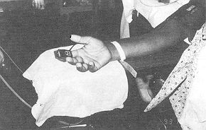
Ashkelon, Israel SUMMARY. llis paper describes a case of finger injury caused by an oxygen saturation monitor probe. A review of the literature is also provided. The patient was subjected to a 7-hour procedure to correct maxillofacial and.nasal deformities, during which oxygen saturation was monitored by means of a nondisposable oximeter sensor placed on the third finger of the right hand. When the probe was removed, a 1.5 x 2 cm blister and a deep circular burn, coinciding with the photo-transmitter side of the probe, were found. The injury was treated conservatively with silver sulphadiazine and healed in 3 weeks. It is recommended that probes should be moved to a different finger every 3 to 4 hours and not be placed too tightly. A case is reported of finger injury by an oxygen saturation monitor probe. Particular attention should be paid in the use of this device during lengthy procedures to avoid risks of pressure necrosis and overheating. Simple precautions which avoid prolonged pressure on the probe and the finger as well as routine maintenance and inspections of the device may reduce the risks. Pulse oximetry is standard practice today for noninvasive oxygenation monitoring. It offers continuous monitoring of arterial haemoglobin saturation on a real-time basis, allowing for early detection of life-threatening hypoxia and aids in optimal patient oxygenation management during anaesthesia, postoperative recovery and intensive care as well as during transport and patient home care. As with many apparatuses that come into prolonged contact with the skin, potential injury can occur. We report a case of digital injury from a pulse oximeter probe, with a review of the literature. Case report A 22-year-old white male patient sought surgical improvement of his facial deformities. The patient had a skeletal class Ill malocclusion, consequent to unilateral cleft lip and palate correction in early childhood. A treatment plan was formulated after a clinical, cephalometric and mounted study model evaluation. The medical history was noncontributory. Physical examination found no contraindication for surgery. Pre-operative laboratory studies, urinalysis and radiograms were all within normal limits. The patient was taken to the operating rom for a Thour procedure to correct his maxillofacial and nasal deformities. The oxygen saturation was monitored by means of a nondisposable oximeter sensor, placed on the third finger of his right hand (Fig. 1), where it remained for the entire procedure.
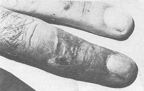
The operative procedure (carried out under hypotensive anaesthetic) included simultaneously: Le Fort 1 osteotomy with advancement and disimpaction, iliac crest bone graft which was inserted between the disimpaction lines, bilateral intraoral vertical mandibular osteotomy, genioplasty and open rhinoplasty. On completion of the operation and removal of the probe, a 1.5 x 2 cm blister and a deep circular burn were noted where the probe contacted the finger, the burn coinciding with the photo-transmitter side of the probe. The injuries were treated conservatively with silver sulphadiazine and healed within 3 weeks, leaving a mild hypertrophic scar (Fig. 2).
Discussion Finger injury associated with a pulse oximeter probe can be caused by one or a combination of several mechanisms. First, the injury may be electrical, resulting from a current path involving the probe and circuitry leaking to the patient through a damaged sensor (Sloan, 1988; Nfiyasaka, 1987). Second, the injury may result from a topical reaction to the probe itself or to residual manufacturing chemicals, Betadine, soap solutions, etc. (Berge, 1988; Chemello, 1990). Third, the injury may result from the mechanical pressure exerted on the finger by the probe. The situation is further compounded by decreased blood flow consequent to controlled hypotension. Tbcking the hand at the side of the patient may have also a role in increasing pressure on the finger (Rubin, 1991; Chemello, 1990). Fourth, overheating of the light-emitting diode (LED) may cause a burn (Sloan, 1988). This last possibility is consistent with the site of the primary injury which corresponds to the region of the LED probe. In our case, we believe the injury was caused by at least two factors, namely pressure and overheating. This may explain the presence of a deep, localized circular burn which exactly fits the LED position and a blister caused by the pressure exerted by the finger clip. Certain precautions should be taken to assure that no morbidity is associated with the use of pulse oximeter sensors. High pressure for short periods or lower pressures for longer periods may cause ischemia. Probes should therefore be moved frequently to decrease the chance of ischemia. The manufacturers recommend routine moving of the probe to a different finger every 3 to 4 hours. There should be frequent checks to make sure the probe is placed accurately on the finger. Tight contact is not required (unlike the use of transcutaneous oxygen electrodes). Probes should be checked for damage and contamination prior to placing on the patient. The region of contact should be inspected for reduced blood flow due to positioning of the probe since this will decrease the removal of the applied heat and increase the likelihood of a thermal burn. These precautions are especially important when controlled hypotensive anaesthesia is applied for a lengthy surgical procedure (e.g. orthognathic procedures). RESUME. Les auteurs décrivent un cas de lésions digitales provoquées par une sonde de monitorage de la saturation de l'oxygène. E y a aussi une revue de la littérature. Le patient a été opéré pour corriger des défauts maxillo-faciaux et nasaux. Pendant l'intervention la saturation de l'oxygène a été contrôlée moyennant un détecteur oxymétrique réutilisable posé sur le troisième doigt de la main droite. A l'enlèvement de la sonde on a trouvé une vésicule de 1,5 x 2 cin et une brûlure profonde circulaire en correspondance de la face photo- transmettante de la sonde. La lésion, traitée en manière conservative avec la sulfadiazine argentée, s'est cicatrisée après trois semaines. Les auteurs recommandent de transférer la sonde à un autre doigt toutes les 3 ou 4 heures et de ne pas l'appliquer trop fort. BIBLIOGRAPHY
|