| Ann. Medit. Burns Club - vol. 6 - n. 3 - September 1993
THE EXTENDED DEEP INFERIOR EPIGASTRIC FLAP: A SPEARHEAD TO
CHARGE INTO THE MOST RESISTANT SITES
Kadry M., Noureldin A.A., Khalifa I.G.
Plastic Surgery Unit, Faculty of Medicine, Cairo University,
Egypt
SUMMARY. The authors present
their experience of the extended deep inferior epigastric flap (EDIEF) for the coverage of
over 30 cases of extensive compound defects of the groin, lower abdomen, perineum and
trochanteric areas, and the thigh, as well as distant areas such as the hand where this
flap can be used both as a distant or as a free flap. The EDIEF, though described since
1983 by Taylor et al., has not been given enough trials for extensive clinical
application. The indications for its use, its limitations, and the technical difficulties
and hazards during use, as well as its application in different ages and sex, are
demonstrated and discussed. The EDIEF seems to be one of the most robust and the most
consistently vascularised flaps. Its length and width, the arc of rotation, the presence
of both muscular and fascial portions, the ability to include other musculocutaneous and
random territories of the epigastric system, its rich vascularity, plus the size of the
flap and of the deep inferior epigastric artery make it one of the most versatile and
reliable flaps for the coverage of large defects.
Introduction
The epigastric vascular arcade
supplies a wide area of the anterior abdominal wall and exhibits a strategic location
providing a highly versatile multicomponent flap that can bridge defects from chest to
knees (Brown et al., 1975). The vascular tree of this part of the body is a link between
the subclavian vessels (via internal mammary and superior epigastric vessels), external
iliac vessels (via inferior epigastric vessels) and intercostals (via costomarginal.
vessels) (Boyd et al., 1984).
These excellent communications between such sizable vessels ensure a highly vascular
territory and hence construction of a safe transfer of various tissue (Corlett and Taylor,
1980). One of the best uses of this vascular arcade is the extended inferior epigastric
flap (ElEF) (Boyd et al., 1984) which is a multicomponent transfer formed of axial
component (inferior epigastric vessels), muscular component (rectus abdominis muscle),
fasciocutaneous component (of vessel dispositions and locations), and a bony component
(9th or 10th rib). The resulting flap is long (nearly 1/4 of body height), narrow based
(trunk of inferior epigastric vessels), and reaches as far as chest, breast, anterior
trunk, perineum, posterior trunk, flank, groin and thigh down to ipsilateral knee (Mixter
et aL, 1989).
Material and Methods
Thirty cases of various defects were treated by using
the E1EF during the period 1989-1992 in the Kasr-El-Aini and AI-Salam Hospitals, Egypt.
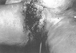
Twelve cases were groin defects, ten of which were contracted scars treated by release
and/or excision, one case was epithelioma of the vulva and groin fold treated by radical
vulvectomy, and the last was a histiocytoma of the groin excised with a safety margin. All
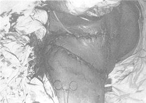
defects were covered using the E1EF and donor site closed primarily.
Five cases of bed sores affecting the trochanteric, iliac crest, and sacral regions were
treated by bursectomy, debridement and covered E1EF. One of these was an axial flap based
on skeletonized inferior epigastric vessels to enable 120' rotation of the flap to cover a
trochanteric ulcer. Four cases of infected traumatic wounds of the femoral triangle were
cleaned and covered by E1EF to save the limbs from risk of secondary haemorrhage. One case
of haemangioendothelioma of the lateral aspect of the thigh, which was excised leaving a
large defect, was successfully closed using the ElEE Three cases of lacerated wounds
around the elbow, with various extents of soft tissue loss and exposure of nerves and
vessels and parts of bones, were treated by debridement and primary neurovascular repair
and covered by the fasciocutanteous element of the flap only, which was detached 3 weeks
later.
Two cases of degloved injury of the forearm were also treated by the same technique. Two
flaps were used simultaneously to cover both hands and distal forearms of a post-burn
contracted scar (PBCS) in a 5-year-old child.One case of above-knee amputation, with
exposure of distal end femur, was covered by the E1EF.
Results
In this work 30 flaps were performed
to demonstrate the clinical applications and versatility of the E1EF. The flap was
successfully used to cover defects of different aetiology and location. One case however
developed severe venous congestion and gangrene, while another suffered loss of the distal
5 cm of the fasciocutaneous component (see Table).
Discussion
It was always been the dream of the
plastic surgeon to find an ideal flap that is highly versatile, safe, with a wide arc of
rotation, able to reach a distant destination, with rich blood supply, easy design, rapid
raising and not bulky. The EIEF seems to top the list of flaps achieving the above
criteria (Boyd et al., 1984).
The anatomical study done by Taylor et al. (1984) showed that the EIEF is nourished by the
deep epigastric system that forms the abdominal portion of a vascular railroad which links
the subclavian and iliac vessels and provides reliable perforators that permit the raising
of several flaps.
In our work we found that a skin flap of large dimensions can be raised in one stage. The
largest was 36 cm. This was extended to the scapular line, although we lost the distal 5
cm of this flap, i.e. nearly up to the post-axillary line. We also succeeded in raising
two other flaps safely to the post-axillary line without any ill effects. The longest flap
done by Taylor et al. (1983) reached only as far as the midaxillary line.
The disc in the anterior rectus sheath, necessary to capture the cutaneous perforators,
was narrowed by carefully approaching the vessels from both medial and lateral directions.
In this way the skin island mobility is increased and hence its arc of rotation. The
narrowing of the disc until it carries two perforators is optimum for flap survival,
although Taylor et al. (1984) harvested only one perforator and luckily enough the flap
survived.
This flap is advantageous in that it has a wide arc of rotation which reaches almost to
the ipsilateral knee and contralateral mid-thigh (Gottlieb et al., 1986). In our work we
were able to cover a defect following soft tissue sarcoma immediately above the knee on
the same side (as done by Gottlieb et al., 1986), to cover a defect over the medial
condyle femur following liposarcoma excision.
Lewis et al. (1980) described the use of a nondelayed thoraco-epigastric flap for a defect
of the upper extremity.
In our work we used the fasciocutaneous element of the EIEF alone to cover the forearm and
hand of a child with a defect following excision and release of a PBCS, with preservation
of most of the integrity of the anterior abdominal wall. The fasciocutaneous element of
the flap proved to be supple, soft, not bulky and is equal to the Chinese flap in many
aspects and even superior in having less donor site morbidity.
The versatility of the flap was demonstrated by Taylor et al. (1984) in 18 patients, when
they used it as a free flap in 15 cases and as a pedicle in three.
In our work we demonstrated the versatility of the flap as a pedicled flap to cover large
defects in the groin, trochanters, forearm and hands, and to reconstruct the vulva and
perineum.
Out of 30 cases we had two complications, one with venous congestion and gangrene, most
probably due to too much bending of the flap, the other with distal 5 cm flap necrosis due
to too much extension of the flap to the scapular line.
The donor defect was successfully closed primarily in all cases without the need to mesh,
and no hernia developed in any of our cases, which were followed up for between some
months to two years post-operatively. They all have an accepted scar. In one case we used
the two sides to provide two flaps simultaneously to cover post-burn defects in both upper
limbs and we closed the donor defects primarily by utilizing the ample tissue reserve
provided by the anterior abdominal wall and saving the patient much suffering by avoiding
a multistaged procedure such as the classical jump flap or some other technique.
With our experience in 30 cases we are convinced that this procedure is safe, speedy and
reliable, and that it fulfils almost all the criteria sought by every reconstructive
surgeon.
 |
 |
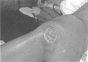
| Fig. (1-A) |
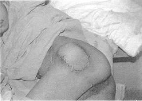
Fig. (I-B) |
 |
 |
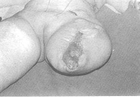
| Fig. (2-A) |
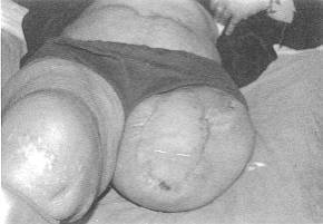
Fig. (2-B) |
 |
 |
| Fig. (3-A) |
Fig. (3-B) |
|
| Site of
Defect |
No. of Cases |
Aetiology |
Type of Flap |
Results |
| Groin |
12 |
10 PBCS |
Pedicled E1EF |
Complete survival |
| |
|
2 malig. tumours |
Pedieled E1EF |
Venous gangrene (one case) |
| Gr. Troch |
1 |
Bed sore |
Pedicled E1EF |
Complete survival |
| 1. crest |
2 |
Bed sore |
Pedicled E1EF |
Complete survival |
| Sacrum |
2 |
Bed sore |
Pedicled E1EF |
Complete survival |
| Fern. triangle |
4 |
Infected wound |
Pedicled E1EF |
Complete survival |
| Lat. thigh |
1 |
Malig. tumours |
Pedicled E1EF |
Complete survival |
| Elbow |
3 |
Trauma |
Pedicled E1EF |
Tip necrosis (one case) |
| Distal UL |
4 |
2 degloved ing. |
Pedicled E1EF |
Complete survival |
| |
|
2 PBCS |
Pedicled EIEF |
Complete survival |
| Amp. kne |
1 |
Pressure sore |
Pedicled E1EF |
Complete survival |
|
|
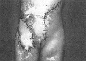 |
Fig. 4 |
|
RESUME. Les auteurs présentent les
résultats de leur expérience de l'emploi du lambeau épigastrique intérieur profond
étendu pour la couverture de 30 cas de défauts composés étendus dans la région de
l'aine, l'abdomen inférieur, le périnée, les trochanters et la cuisse, comme aussi dans
les régions distantes comme la main, où ce type de lambeau peut être utilisé soit
comme lambeau à distance soit comme lambeau libre. Le lambeau épigastrique inférieur
profond étendu a été décrit pour la première fois par Taylor et coll. (1983) mais il
a eu une application clinique très limitée à cause de l'insuffisance des épreuves
pratiques. Ici les auteurs donnent des indications pour ce qui concerne son emploi, ses
limitations, les difficultés et les dangers techniques pendant l'emploi, et l'application
chez les patients masculins et féminins d'àge divers. Ce type de lambeau parait être un
des lambeaux les plus résistants et les plus régulièrement vascularisés. Sa Ion ueur
et sa largeur, l'arc de rotation, la présence de portions non seulement musculaires mais
aussi fasciales, la capacité d'embrasser 9 d'autres territoires musculocutanés et
variés du systyme épigastrique, sa riche vascularité, les dimensions du lambeau et de
l'artère épigastrique inférieure profonde - tous ces facteurs se combinent pour faire
de ce lambeau une des méthodes les plus universelles et fiables pour la couverture des
gros défauts cutanés.
BIBLIOGRAPHY
- Boyd J.B., Taylor GI Corlett R.J.: The vascular
territories of the superior epigastric and the deep inferior epigastric systems. Plast.
Reconstr. Surg., 73: 1, 1984.
- Brown R.G., Vasconez L.O., Jurkiewiez M.L:
Transverse abdominal flaps and the deep epigastric arcade. Plast. Reconstr. Surg., 55:
416, 1975.
- Corlett R.J., Taylor GI: The angiotomes of the body
and their relation to local and distant tissue transfer. Paper presented at the British
Association of Plastic Surgeons Summer MeetiDg, July 1980.
- Gottlieb M.E. et al.: Clinical applications of the
extended deep inferior epigastric flap. Plast. Reconstr. Surg., 78: 782, 1986.
- Lewis V.L. Cook J.Q.: The nondelayed
thoracoepigastric flap: coverage of an extensive electric burn defect of the upper
extremity. Plast. Reconstr. Surg., 65: 492, 1980.
- Mixter R.C., Wood W.A., Dibbell D.G.:
Retroperitoneal transposition of rectus abdominis myocutaneous flaps to the perineum and
back. Plast. Reconstr. Surg., 65: 437, 1990.
- Taylor G.I. et al.: The extended deep inferior
epigastric flap: a clinical technique. Plast. Reconstr. Surg., 72: 751, 1983.
- Taylor G.I. et al.: The versatile deep inferior
epigastric (inferior rectus abdominis) flap. Brit. Plast. Surg., 37: 330, 1984.
|