| Ann. Medit. Burns Club - vol. V1 - n. 4 - December 1993
THE PHYSICOCHEMICAL CHARACTERISTICS OF KELOID COLLAGEN BEFORE AND AFTER CLINICAL TREATMENT WITH KENACORM A POLARIZING MICROSCOPICAL STUDY Chavrakov W.*, Mazgalova J.** * Clinical Centre of Endocrinology and
Gerontology, Ivan Penchev University Hospital, Department of Pathology, SUMMARY. Collagen structures of the keloids were studied by polarized light microscopy. The purpose of the investigation was to determine the histophysical characteristics of the collagenous fibrils before and after clinical treatment of keloids with Kenacort. The method of imbibition analysis was used. The total anisotropy of keloid collagen after the cure was measured and compared with the anisotropy of the collagen before treatment. An assessment of the rate of collagen rehydration was made by using total birefringence rates. It was found that the rate of collagen rehydration was faster in the group of treated keloids than in untreated lesions. Data for the histophysical state of the collagen were established. It is suggested that the variation of collagen rehydration could be explained by the alterations in the mucopolysaccharides of the ground substance. Two hypotheses for the explanation are put forward. Formation of keloids over a period of many
years is among the most common healing problems in patients who survive thermal injury.
This type of bum complication, besides its significance as a cosmetic deformation, may be
a cause of different functional disorders in the organism. This fact has drawn the
interest of researchers to the unexplained moments of the mechanism of keloid formation
and to the more or less successful efforts for their cure. The special literature offers
detailed information regarding the histiotypic organization of keloids and hypertrophic
scars and their specificity, after examination by scanning and transmission electron
microscopy (4, 1, 2, 3). However, data on the morphological changes of the structural
elements of the keloids, as treated in various manners, are scant. There are no reports on
the physicochemical characteristics of these keloids. Materials and methods Steroid treatment on keloids in
patient~volunteers was performed at the Pirogov Emergency Institute, Section of Bums and
Plastic Surgery, Sofia, Bulgaria. Subjects from either sex in the age range of 12 to 50 yr
were subjected to treatment with Kenacort. The keloids were treated with an average dose
of 20 mg/ml triamcinolone acetate, applied sublesionally. All details of clinical
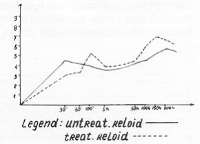
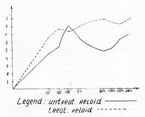
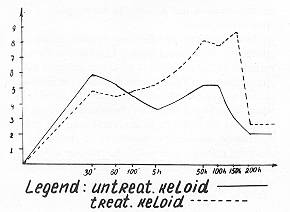
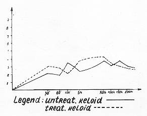
treatment will be published in another report. R = I + F (8). It should be noted that total anisotropy of the collagenous fibre is also a function of the cross diameter of the fibre which in contemporary studies is measured by interference microscopy. For this reason the results of our investigations for the measuring of birefringence have only a relative value. All tissues were obtained from surgical excisions. Every keloid was cut into 5 small pieces for examination. Sixteen microscope slides were prepared for the investigation with each hydrophilic medium. The anisotropy rates given in Table I are an average of the anisotropy rates of 10 collagenous fibres per slide. Total anisotropy was calculated in nanometers by Wiener's formula (6) and presented with the meaning of X for every group and every interval of time. Data were elaborated statistically with the finding of t and R Results The data for the total anisotropy of keloid collagen before and after clinical treatment, depending on the time intervals and the type of incubation medium, are shown in Table I.
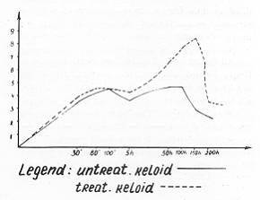
.On the basis of these, the curves for the velocity of collagen rehydration in different immersion media are built. An assessment of the velocity is made by means of the rates of total birefringence in nanometers. It is clear that the velocity of collagen rehydration is faster in the group of treated keloids than in the group of untreated lesions in most of the investigative time intervals investigated. The rate velocity is highest in the group of treated keloids after 50 h (Figs. 2, 3, 4 and 5) or at exactly the fiftieth hour (Fig. 1). The curves show a diminution in the anisotropy rates after this peak without any further increase. The rates of bireftingence during incubation with water show a new increase after 150 h (Fig. 2). In the group of untreated keloids the curves demonstrate the variability of the rate of collagen rehydration. There are some considerable rises and reductions in the rates of anisotropy which cause changes of inconstant type (Figs. 1, 2, 3, 4 and 5). The distinctions between the rates of the two groups are statistically reliable (Table 1).
Discussion Data regarding the histophysical state
of keloid collagen before and after clinical treatment are considered. The possibilities
of polarizing microscopy were used to gather indirect information on the molecular
organization, which defines the interrelations between the structural elements of collagen
and the other components of the connective tissue. Total anisotropy of the collagenous
fibres was used as one of the indices for the estimation of keloidal collagen. The
imbibition curves gave information only regarding the velocity of collagen rehydration in
different immersion media, as estimated by the variable rates of bireffingence. On the
basis of these results we tried to discover whether the keloidal collagenous fibrils are
rehydratable and whether there are any differences in their rehydration rates before and
after clinical treatment.
RESUME. Les auteurs ont étudié les structures collagènes moyennant la microscopie polarisante. Le but de l'investigation était de déterminer les charactéristiques histophysiques des fibrilles collagènes avant et après le traitement clinique des chéloïdes avec Kenacort. En utilisant la méthode de l'analyse de l'imbibition, ils ont mesuré l'anisotropie totale du collagène chéloïde après le traitement et ils l'ont confrontée avec l'anisotropie du collagène avant le traitement. En outre ils ont calculé la vélocité de la réhydratation collagène moyennant les vélocités de la biréfringence. Ils ont trouvé que la vélocité de la réhydratation collagène était majeure dans le groupe des chéloïdes traités par rapport aux lésions non traitées. Les données pour l'état histophysique du collagène ont été établies. Les auteurs proposent que la variation de la réhydratation collagène pourrait être causée par les altérations dans les mucopolysaccharides de la substance interstitielle, et ils avancent deux hypothèses pour justifier cette possibilité. BIBLIOGRAPHY
|
|||||||||||||||||||||||||||||||||||||||||||||||||||||||||||||||||||||||||||||||||||||||||||||||||||||||||||||||||||||||||||||||||||||||||||||||||||||||||||||||||||||||||||||||||||||||||||||||||||||||||||||||||||||||||||||||||||||||||||||||||||||||||||||||||||||||||||||||||||||||||||||||||||||||||||||||||||||||||||||||||||||||||||||||||||||||||||