Ann. Medit Burns Club - voL VII - n. I - March 1994

THE USE OF TISSUE EXPANSION IN BURNS SEQUELAE

Pérez Barrero P., Duato F., Agullô A., Gonzàles Labasa M., Vistôs J.L., Cimorra G.A.

Plastic and Reconstructive Surgery Department and Regional Burns Unit, Hospital Miguel Servet, Zaragoza, Spain


SUMMARY. Tissue expansion has become a very useful tool in the reconstruction of aesthetic and functional burns sequelae. In this paper we review a series of 56 expansions in 44 patients with burns sequelae treated in our department. We describe the technique and complications. According to our experience, the use of soft-tissue expansion for the reconstruction of scarred deformities in burn patients has proved to be a technique with optimal aesthetic and functional results. We therefore believe that controlled tissue expansion should be considered an extremely useful technique in the burn patient and should he selected when considered as an alternative.

Controlled tissue expansion is a very worthwhile tool for the plastic surgeon who approaches the reconstruction of burns sequelae by covering the defects with adjacent donor tissues of similar colour, innervation, hair-bearing qualities and texture, leaving intact the adjacent skin structures. This technique has the unique characteristic of providing us with extra tissue without leaving an unsightly donor site.The sequelae of most burn wounds involve a certain degree of contracture, even if wound healing is by epithclialization of a graft. To have a shortage of donor tissue with similar characteristics is almost the rule. Furthermore, when we are planning the correction of the resulting deformities we should consider that the real defect is usually greater than the apparent one.The increase in vascularity produced during the expansion enables us to produce larger, reliable and predictable flaps. It also makes it easier to close donor sites.This technique therefore offers new prospects for treating burns sequelae because it allows us to increase the amount of surrounding non-affected skin and to use it to cover the excision of scarring wounds by local or distant flaps.In the light of present-day experience, the technique is now available to all plastic reconstruction surgeons. Like any other technique tissue expansion has some disadvantages, including the necessity of two - or even more - surgical procedures as well as multiple visits to the out-patients clinic. There is also a certain discomfort while expansion is taking place, plus a transitory cosmetic defect as the expanders become larger and therefore highly visible.

Material and methods

All patients with tissue expanders placed between 1988 and 1992 in our Service were reviewed. In this 5-year period, we placed 56 expanders in 44 patients who had suffered burns sequelae.
The femalelmale ratio was 25 to 15 (62 to 38%). The ages ranged from 3 to 44 yr with a mean age at surgery of 17 yr.
We used 56 expanders in different forms and sizes. The maximum volume filled oscillated between 45 and 800 cc, depending on the site.

Head and neck


35

  (Face          15 Scalp                  14 Neck              6)

Trunk


8

  (Breast         2 Abdomen             6)

4

Upper limb


5

Lower limb


4

Table 1 - Number of expanders by regions

Two expanders were used simultaneously in nine patients (5 in the scalp, 2 in the face, 2 in the lower limbs). In another patient we used four expanders in the trunk.
Table I shows the number of expanders by regions.Every one of these areas needs specific tissue requirements and accurate instructions for its reconstruction.

Surgical technique

Pre-operative counselling with the patients is essential. They must be aware that cosmetic deformities will occur during expansion; that at least two surgical procedures will be required; and that they could suffer mild discomfort during expansion. An informed and well-motivated patient will tolerate this much better.
The design of the flap has to be well studied preoperatively. It is mandatory to avoid scarred, atrophied or radiated areas. However, in breast reconstruction, it is impossible not to enclose the scar in the expansion. When a correct indication has been established an important decision has to be taken: the location of the expander and the remote filling port.
The most important decision is the placing of the incision: this must be short and as distant from the defect as possible.Incisions radial or perpendicular to the defect are preferable.The optimal localization of the filling port (unless we use the filling port included in the expander) is in a silent area above or by the side of the expander.A good way to estimate the length of the flap to be created is to double the distance from the skin surface to the dome of the expander and then subtract the length of the base.An over-expansion of 30 to 50% is a safe procedure, because it allows the production of a larger amount of tissue.
Large defects will demand multiple peripheral expanders. The best results are obtained with expanders of larger volume than first estimated.In our cases we used only expanders with a remote filling port.We do not give prophylactic antibiotics as a routine measure.

First stage

The patient is subjected to general endotracheal anaesthesia, through a conveniently placed incision, and a subcutaneous pocket is dissected (subgaleal in the scalp). The size of the pocket must be sufficient to accommodate the temporary implant, which must remain with a smooth surface filling it adequately.
The intra-operative filling of the expander reduces the need of drains to prevent the formation of haematomas and seromas. It also reduces the formation of folds on the expander surface which could eventually damage the skin.
In the scalp, care must be taken with the use of electrocautery to prevent burning of the hair follicles.The remote filling port must be located in an adjacent pocket that is however well apart from the expander, preferably over a bony surface, in order to facilitate its localization at the filling phase. The air is aspirated from the expander and a variable amount of saline is injected, in sufficient quantity to allow easy closure of the incision and a smooth surface.

Scalp

7 weeks (5-12)

Face

7 weeks (5-11)

Neck

8 weeks (5~10)

Trunk

11 weeks (7-13)

Upper limb

9 weeks (6-10)

Lower limb

9 weeks (6-11)

Table II - Expansion-time average by regions

Implant inflation

We begin expansion two to four weeks after the incision of the implant placement has healed. For successive inflations we use saline with the addition of a certain quantity of dye (Bonney's blue) to facilitate location of the filling port. The amount of fluid injected each time is not subject to any rule: it is individualized according to the degree of tension and blanching of the skin, and to patient comfort. The filling sessions are usually at one-week intervals. In our series the expansion time ranged according to the region (Table II).

Fig. 1A - Expander located in mandibular region in 17-year-old girl presenting a burn in the cheek area. Fig. 1A - Expander located in mandibular region in 17-year-old girl presenting a burn in the cheek area.

Second stage

When the planned expansion has been completely achieved, the patient is re-admitted one day before the next surgical procedure.

With the patient under general anaesthesia the expander is removed, the flap redesigned and the defect excised in accordance with the available tissue. This sequence is important in order to avoid creating an excised defect and a flap with insufficient skin to cover it. We do not make capsulotomies, in order to avoid damage to the neovascular net which grows around the capsule.

Fig. 1B - Early post-operative result. Fig. 1B - Early post-operative result.

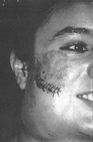
One-layer closure is suitable for the scalp. In other regions the suturing is performed in two layers. In this second phase we do not use any solution containing adrenalin to reduce bleeding, since it has been proved experimentally that the addition of this substance, even in a proportion of 1 to 200,000, markedly decreases the survival of delayed flaps.

Case 1

Girl, 17 years old, with 55-mm-diameter scar over the right cheek-bone where, three months earlier, she had suffered a deep contact burn which was debrided early and covered with mid-thickness Wolfe-Kraus grafts. We planned a rotation-advancement flap from the adjacent skin of the cheek, placing a 200 mI expander (Fig. ]A) under it in order to get sufficient cutis. We proceeded with the expansion and ten weeks later excised the scar and covered it with the expanded flap. There were no complications and the result was acceptable (Fig. ]B).

Fig. 2A - Post-burn alopecia and expander over the parietooccipital region in 5-year-old boy. Fig. 2B - The alopecia area rerrioved and the flap raised.
Fig. 2C - Early post-operative result. Fig. 2C - Early post-operative result.

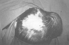
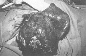
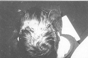
Case 2

Child, aged 5 years, showing post-burn alopecia 100 mm in diameter over the right parietal region due to an injury sustained two years before. We designed a rotation flap to cover the defect and expanded it with 150 ral placed subgaleally over the parieto-occipital region (Fig. 2A). After 9 weeks' expansion we removed the alopecia (Fig. 2B) and covered the area, advancing the expanded flap (Fig. 2C).

Fig. 3A - Burn scArs over both. thighs in 19-year-old girl. Fig. 3B - Right thigh with the expancler.
Fig. 3C - Early post-operative resuit, right thigh. Fig. 3D - Early post-operative result, lelt thigh.
Fig. 3C - Early post-operative resuit, right thigh.

Fig. 3D - Early post-operative result, lelt thigh.

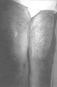
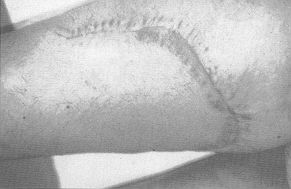
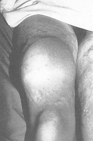
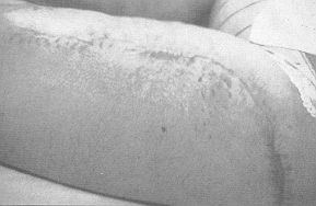
Case 3

Long-lasting scars over the anterior and inner face of both thighs in a 19-year-old girl who had suffered deep dermal burns 9 years previously (Fig. 3A). We used two expanders, 700 mI on the right leg (Fig. 3B) and 640 mI on the left. Nine weeks later we excised the scars and advanced the flaps covering the defects (Figs. 3C and 3D) without any post-operative complication.

Fig. 4A - Large post-burn scar in trunk of 15-year-old girl. Fig. 4B - The four expanders used.

Fig. 4A - Large post-burn scar in trunk of 15-year-old girl.

Fig. 4B - The four expanders used.
Fig. 4C - Early post-operative result, pending right breast construction. Fig. 4C - Early post-operative result, pending right breast construction.

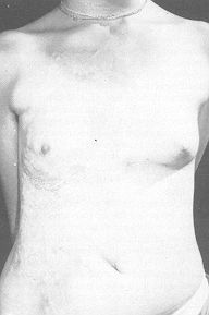
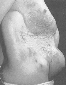
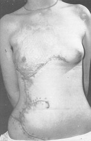
Case 4

Large post-burn scar on the trunk of a girl aged 15 years affected by thermal lesions when she was a child (Fig. 4A). Four large expanders were used, placed around the defect, and the period of inflation was 11 weeks (Fig. 4B). The result is shown in Fig. 4C. We are waiting for the girl to grow a little more before carrying out a right breast reconstruction procedure.

Results

Fifty-six expanders were used, with complications in just 6 cases (10%). The complications were classified as major or minor. The major complications were those that required removal of the expander or a change in the initial planning. Three cases fell into this category (5%). Two of these (both in the anterior cervical area) necessitated removal of the expander because of exteriorization. In the third case (expander under the scalp), expander dislocation occurred, making resettling necessary. In three cases (5%) minor complications were present: two patients suffered a small suture dehiscence (one in the breast and one in the scalp); the third patient, with a face expander, had a haematoma which was drained without further problems.
The patients showed a high degree of satisfaction. In most cases the aesthetic results were very good.

Discussion

When we are faced with the reconstruction of postburn sequelae, we usually find a shortage of neighbouring donor tissues with similar tissue properties. Furthermore, the real size of the actual defect after excision of the scar is larger than that we see. This is due to the contraction forces produced in every wound healed by second intention or with the use of thin autologous skin grafts.
Tissue expansion has clearly increased our possibilities for dealing with burn reconstruction problems. Nevertheless, the introduction of a relatively new and very useful technique must not affect the need for judicious preoperative evaluation. It is also important to ensure a careful surgical technique consistent with all surgical principles.
The preferential use of radial incisions will diminish the risk of wound dehiscence and therefore shorten the expansion period.
Expanded rotation flaps usually give better results than advancing flaps. Considering the morbidity of donor areas, secondary defects, etc., the results obtained with this technique are, in our opinion, better than those we can achieve with other traditional methods such as partial- or full-thickness skin grafts, distant flaps or free-flaps. These techniques can have particular applications in the burn patient.
It is very important to consider that major complications cause only a delay in the planned reconstruction with no loss of additional tissues in most cases.
The complication rate in our series is low: the expanders had to be removed in only two cases. In all the rest the planned reconstruction was accomplished successfully.
In conclusion, according to our experience, the use of soft-tissue expansion for the reconstruction of scarred deformities in the burn patient has proved to be a technique with optimal aesthetic and functional results. We have achieved a high degree of patient satisfaction with the repairs. We therefore believe that controlled tissue expansion should be regarded as an extremely useful technique in the burn patient and should be preferably elected when considered as an alternative.

RESUME. L'expansion tissulaire s'est montrée une technique très utile dans la reconstruction des séquelles esthétiques et fonctionnelles des brûlures. Cet article considère une série de 56 expansions chez 44 patients atteints de séquelles de brûlures traités dans notre service.
Nous décrivons la technique et les complications. Selon notre expérience l'emploi de l'expansion du tissu mou pour la reconstruction des difformités cicatrisées du patient brûlé s'est montré une technique qui offre des résultats esthétiques et fonctionnels excellents. A notre avis, par conséquent, l'expansion tissulaire contrôlée est une technique extrêmement utile pour le patient brûlé qu'il faut préférer dans le cas où il y a des alternatives possibles.


BIBLIOGRAPHY

  1. Adson M.: Scalp expansion in the treatment of male pattern baldness. Plast. Reconstr. Surg., 79: 906, 1987.
  2. Antonyshyn 0., Gruss J.S., Zuker R., MacKinnon S.E.: Tissue expansion in head and neck reconstruction. Plast. Reconstr. Surg.,82:58,1987.
  3. Argenta L.C., Watanabe M.J., Grabb W.C.: The use of tissue expansion in head and neck reconstruction. Ann. Plast. Surg., 11: 3 1,1983.
  4. Argenta L.C.: Controlled tissue expansion. Br. J. Plast. Surg., 37: 520,1984.
  5. Argenta L.: Advances in tissue expansion. Clin. Plast. Surg., 12:159, 1985.
  6. Argenta L.: Principles and techniques of tissue expansion. In: McCarthy: "Plastic Surgery", W.B. Saunders Company, Philadelphia, 1990.
  7. Austad E.: Tissue expansion: dividend or loan. Plast. Reconstr. Surg., 78: 63, 1986.
  8. Baux S., Mimoun M., Hilligot P.: Cutaneous expansion in burns sequelae. Chirurgie, 116: 373, 1990.
  9. Cherry G.: Increased survival and vascularity of random-pattern skin flaps elevated in controlled expanded skin. Plast. Reconstr. Surg., 72:680,1983.
  10. Leighton W.D., Johnson M.L., Friedland J.A.: Use of the temporary soft-tissue expander in post-traumatic alopecia. Plast. Reconstr. Surg., 77: 737, 1986.
  11. Leonard A.G., Small J.O.: Tissue expansion in the treatment of alopecia. Br. J. Plast. Surg., 39: 42, 1986
  12. Manders E., Schenden M.J., Furrey J.A., Heztler P.T., Davis T.S., Graham W.P.: Soft-tissue expansion: concepts and complications. Plast. Reconstr. Surg., 74: 493, 1984.
  13. Manders E.: Soft-tissue expansion in the lower extremities. Plast. Reconstr. Surg., 81: 208, 1988.
  14. Neale H.: Complications of controlled tissue expansion in the pediatric burn patient. Plast. Reconstr. Surg., 82: 840, 1988.
  15. Neumann C.: The expansion of an area of skin by progressi\e distension of the subcutaneous balloon. Plast. Reconstr. Surg., 19. 124,1957.
  16. Radovan C.: Breast reconstruction after mastectomy using the temporary expander. Plast. Reconstr. Surg., 69: 195, 1982.
  17. Radovan C.: Tissue expansion in soft-tissue reconstruction. Plast. Reconstr. Surg., 74: 482, 1984.
  18. Sasaki G.H., Pamg C.Y.: Pathophysiology of skin flaps raised on expanded pig skin. Plast. Reconstr. Surg., 74: 59, 1984.
  19. Spence R.J.: Experience with novel uses of tissue expander in burn reconstruction of the face and neck. Ann. Plast. Surg., 28: 453-64, 1992.
  20. Van Rappard J.: Surface-area increase in tissue expansion. Plast. Reconstr. Surg., 82: 833, 1988.
  21. Wieslander J.B.: Tissue expansion in head and neck. A 6-year review. Scand. J. Plast. Reconstr. Surg., 25: 47-56, 1991.
  22. Zellweger G.: Tissue expansion in reconstruction of burn sequelae. Ann. Plast. Surg., 26: 380-8, 1991.


Contact Us
mbcpa@medbc.com