| Ann. Medit. Burns Club - vol. VII - n. 3 - September 1994
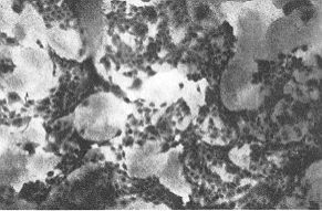
HEALING EFFECT OF RECOMBINED HUMAN/PIG SKIN ON DERMAL DEFECTS Matou~kovd E, Ndmcovd D., Dvofdnkovd D., VogtovA D., KénigovA R. Institute of Molecular Genetics AV CR, Prague SUMMARY. The healing effect of recombined human/pig skin (R11PS), composed of human keratinocytes growing on cell-free xenodermis, was studied. When R11PS was applied with the keratinocyte layer facing the wound, stimulation of healing of leg ulcers and deep-dermal wounds was observed. The structure of RHPS makes manipulation easier and more effective compared to cultured epidermal sheets. Introduction As autologous skin is not available in massive burns and is often wasted in chronic skin defects, surgeons are still searching for an ideal skin substitute (1, 2). Cultured epidermis has been used to treat skin loss since 1981 (3), but a satisfactory dermo-epidermal skin substitute that is readily available in suf~ ficient quantities has still to be found. On the basis of long experience with the application of xenografts in bum treatment (4) and of experience with cultured epiderinis (5, 6) we have developed recombined human/pig skin (RBPS), a composite skin substitute which consists of human autologous or allogeneic keratinocytes cultivated on cell-free pig dermis (7). In this work we describe the healing effect of RBPS containing allogeneic keratinocytes on chronic leg ulcers and bums. Methods Short description of the method of RHPS preparation (for details see Bibliography, 7) Epidermis and fibroblasts are removed from strips of sterile, living pig skin after trypsinization. The resulting dermis is macerated in water (to wash out the rest of the cells, salts and antibiotics), spread on the tissue culture dish and stuck to it by drying, forming a thin collagen film. Dry dermis behaves as a firm substrate for cell culture. The dishes containing dried dermis may be steril-ized with gamma-irradiation (not necessary when treated with antibiotics and fungicides) and preserved at room temperature. The dermis is pre-washed with the growth medium for several hours. Lethally irradiated 3T3 cells are then seeded on it in a fresh medium. Keratinocytes (2nd passage) are added the next day. This -method, based on the method of Rheinwald and Green (8), enables us to obtain more than 500 sq cm of RHPS from one sq cm of human split-skin graft. The 3T3 cells can be selectively washed off at any time of culture by differential trypsinization (Fig. 1). Application (?f RHPS
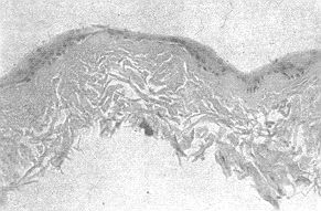
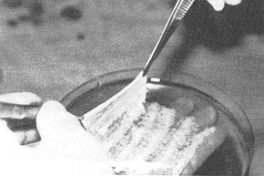
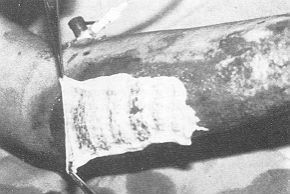
At keratinocyte subconfluency or confluency, usually 711 days after seeding, the recombined skin (Fig. 2), which is very similar in consistency to normal skin, was peeled off without enzymatic release (Fig. 3) and placed on the skin defect (Fig. 4) with the keratinocyte layer facing the wound. It was then covered with a layer of vaseline gauze and several layers of gauze wetted with cholera-toxin-free (in the case of bums also serum-free) growth medium. The RBPS-covered defect was checked two to five days after application. Results Chronic leg ulcers For treatment of leg ulcers RFIPS containing allogeneic keratinocytes was used. The healing effect depended on the size and cleanness of the wound.
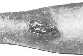
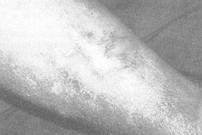
The greatest obstacle to RBPS "take" was wound infection. The most successful method of leg ulcer clean~ing and disinfection was provided by the use of 5% salicyl acid cream. Case 1 R11PS with a subconfluent keratinocyte layer was applied on a leg ulcer 12 sq em in size (Fig. 5a). Rapid healing started from the edges of the wound after three days. The ulcer healed in two weeks and remained in a stable state (Fig. 5b). Case 2 RHPS was applied on a clean 4 sq em deep leg ulcer reaching to the bone. The wound filled up after seven days and only a narrow gap remained on the surface. Burns: RHPS was applied in four patients. Dry application to the donor site was not successful (wetting is necessary). RHPS containing autologous and/or allogeneic keratinocytes applied in a patient with massive burns (80%) was dissolved.
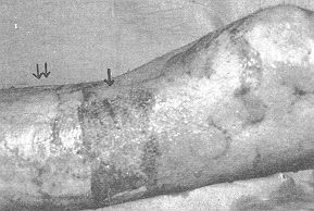
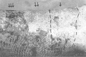
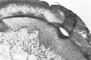
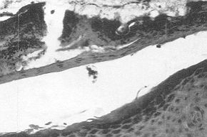
Case 3 A 20-year-old male with deep dermal burns on the leg (I I% BSA) was necrectomized and xenografted five days post-burn. Two days after necrectomy a 5 x 6 cm area of the wound was covered with RHPS containing allogeneic keratinocytes and wetted with the culture medium. The rest of the wound was cover-ed with xenografts. Two days later the xenografts were replaced by autografts, while RHPS remained untouched. Five days after RHPS application RHPS "take" and epithelialization of the treated area were apparent (Fig. 6a). Seven days after RHPS application a histological sample was taken from the RHPS grafted area (Fig. 7a). Dry RHPS started to peel off (Fig. 7b), but the wound underneath healed in two weeks (Fig. 6b). Case 4 A burn wound 60 sq cm in size was covered with R11PS containing allogeneic keratinocytes. In the course of five days 40% of the surface was epithelialized while the rest had to be autografted, probably because it did not contain any basal epidermal cells. Discussion RHPS is a new type of skin substitute,
combining the advantages of xenografts and cultured epidermal sheets. The antigenicity of
pig skin is minimized by removing all potentially antigeneic elements (i.e. epidermis and
other skin cells). Only a collagen matrix preserving the natural structure of the dermis
is used as a firm substrate (dried and stuck to the bottom of the tissue culture dish) for
keratinocyte culture. With the method used in this study - human keratinocytes cultured on
cell-free pig dermis, applied with the keratinocyte layer facing the wound - RHPS had a
remarkable healing action on skin defects. It also stimulated granulation tissue formation
and the filling up of the base of small deep ulcers. When applied in a normal orientation
(i.e. keratinocytes facing upwards), RHPS usually did not "take".
We are currently investigating the development of new generations of RHPS containing fibroblasts incorpo.rated in the dermal structure.
RESUME. Les auteurs ont étudié l'effet guérissant de la peau humaine/porcine recombinée (sigle anglais: RHPS), composée de kératinocytes humains qui croissaient sur le xénoderme libre de cellules. Quand la RHPS était appliquée avec la couche de kératinocyte face à la lésion il était possible d'observer la stimulation de la guérison des ulcères de la jambe et des brûlures du derme profond. La structure de la RHPS rend la manipulation plus facile et plus efficace par rapport aux lambeaux épidermiques cultivés. BIBLIOGRAPHY
|