| Ann. Medit. Burns Club - vol. VII - n. 4 - December 1994
USE OF AMNIOTIC MEMBRANES AS BIOLOGICAL DRESSINGS IN CONTEMPORARY TREATMENT OF BURNS Atanassov W, Mazgalova J.**, Todorov R*, Stereva K***, Trencheva W* * Section of Thermal injury and Plastic Surgery, SUMMARY. The positive results of burn treatment with amniotic membranes are described. The indications for the clinical use of amnion are defined. It is used in promoting early epithelial regeneration and it reduces scar hypertrophy, Introduction The role of amniotic membranes as biological dressings in the treatment of partial-thickness burns is well established (1, 2, 4, 5, 7). Positive clinical results in their application have been reported. The 'results are accompanied by reduction of pain, decreased bacterial count in the burn wound and promotion of epithelialization (2, 3, 6). The use of amniotic membranes causes fewer complications than synthetic skin covers (4, 8). Materials and methods Foetal membranes were obtained fresh
from deliveries (seronegative to syphilis, hepatitis B and AIDS). The chorion was removed.
The isolated amnion was washed with saline, stored in bags containing 70' ethyl alcohol
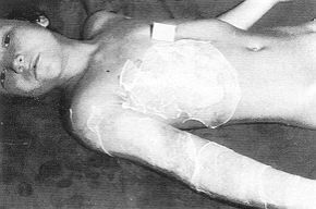
and kept at room temperature. Before use, the amnion was washed in sterile saline. Results The results were observed in patients between 5 and 20 days after the first covering of the wounds with amnion. Nine of the patients were treated on admission to the clinic, six on the second or third day after their thermal injury, five children were placed on Klinitron beds and three in air-therapeutic conditions. The amniotic dressings were not covered in six patients.
The results of the application of amniotic
membranes are as follows:
2. Different-depth dermal burns were
treated with amnion. Burned skin covered with amnion during the first 24 h after the
trauma kept its surface dry for up to seven days. This is very important for wounds on the
back of the body. Amnion is no obstacle to the removal of necrosis or to the application
of auto- and allografts. Its use during the first three days after the burn prepares the
injured area for early tangential necrectomy. When early necrectomy was contraindicated,
the elimination o,f necrosis was completed by chemical necrectomy through the amniotic
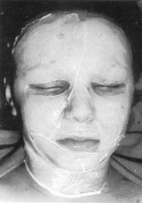
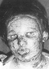
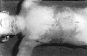
membranes. A well-defined young epithelium was seen when the dried amnion was removed (Figs.
4, 5).
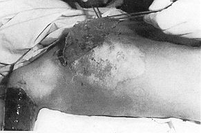
4. Amniotic membranes were used for the
treatment of skin donor sides. When amnion was meshed, coagula were formed and the
dressings were removed. Epithelialization was observed within two weeks when the
mesh-graft dermatom perforated the amnion-covered donor site. Donor wounds treated in this
manner caused less pain, and infection was reduced in comparison to conventional clinical
dressings. Discussion Amniotic membranes have been used as a biological dressing in the treatment of burns of different dermal depth. We returned to this kind of wound cover because of its easy availability and low cost. We try here to define some of the indications for the clinical use of amnion.
Our results clearly show the usefulness of the early application of amniotic membranes in the treatment of fresh superficial bums. Large injured areas were treated by amnion during the first 24 h after injury. With air-conditioning, amniotic membranes remained dry for a week. Their application was not an obstacle to removal of necrosis. We observed a healthy effect of amnion when used for meshgraft covering. We consider it to be effective in view of the promotion of epithelialization, reduction of pain and limited infection. Amniotic membranes are economical and easily available. Amnion is not "taken", it only acts as a protective biological dressing. It does not survive on dead tissue and dissolves on granulating areas. It is useful in promoting the early regeneration of epithelium and reduces scar hypertrophy. RESUME. Les auteurs décrivent les résultats positifs obtenus dans le traitement des brûlures avec les membranes amniotiques. Ils définis sent les indications pour l'emploi clinique de l'amnios, qui est utilisé pour promouvoir la régénération épithéliale précoce. En outre il réduit l'hypertrophie cicatricielle. BIBLIOGRAPHY
EUROPEAN BURNS ASSOCIATION 6TH
INTERNATIONAL CONGRESS The scientific programme includes: Burns in the elderly Scientific Secretariat: Prof. Dino Barisom, Divisione di Chirurgia
Plastica la Meeting Venue: Exhibition Centre Organizing Secretariat: Errebi Congressi dept. of Renbel Travel s.r.l. |