| Ann. Medit. Burns Club - vol. VIII - n. I - March 1995
A CASE OF TOXIC EPIDERMAL NECROLYSES ASSOCIATED WITH
MYCOSIS FUNGOIDES AND COMPLICATED BY CONSUMPTION COAGULOPATHY
Napoli B., D'Arpa N., Sferrazza-Papa G, Masellis M.
Divisione di Chirurgia Plastica e Terapia delle
Ustioni, Ospedale Civico e Benfratelli, USIL 58, Palermo, Italia
SUMMARY. A
case of drug-related toxic epidermal necrolysis (TEN) is described in an adult patient
suffering from mycosis fungoides. The course of the disease, which affected 50% of the
body surface and ended fatally following cerebral haemorrhage, was characterized by the
onset of a serious hepatopathy, thrornbocytopenia and consumption coagulopathy. These
alterations were considered to be TEN-specific and not secondary to the sepsis
subsequently observed. Stress is laid on the complexity of this disease, which is not only
cutaneous or cutaneoushnucoseal, and on the importance of immediate hospitalization in a
specialized centre in order to guarantee continuity of therapeutic treatment.
Introduction
The characteristic skin involvement
makes toxic epidermal necrolysis (TEN) resemble a second-degree bum, with which it has
several physiopathological aspects in common (e.g. loss of liquids, hypereatabolism and
increased infective risk).
TEN is a reaction to drugs which manifests itself not only at the level of the skin but
also in the mucous membranes of various orifices and in a number of organs and systems,
sometimes primarily.
Because of this multisystemic involvement, frequently with hepatic, haematological and
pulmonary alterations, TEN has a worse prognosis than a burn of the same extent. We
describe here a patient suffering from TEN whom we recently observed. The patient died
when skin reepithelialization was to a large extent complete.
Clinical case
S. G., aged 50 years, male
On 20 April 1992, because of intense
bilateral gonalgia, and following a medical prescription, the patient had intramuscular
injections of three phials of a piroxicambased product. Three days later he noticed the
appearance of widespread, non-pruritic, confluent erythernatous patches. On 24 April 1992
he was admitted to the Palermo University Dermatological Clinic.
The patient was given systemic and topical cortisone treatment for six days and then
transferred to the Intensive Care Unit (ICU) on 30 April following the onset of severe
hydroelectrolytic and metabolic imbalance, after the appearance of the typical TEN skin
picture.
In the ICU the patient's hydroelectrolytic equilibrium was restored and he was subjected
to hyperbaric oxygenotherapy sessions, antibiotic and cortisone treatment, and total
parenteral nutrition (TPN) by a central venous route (right vena subelavia).
When the patient reached our Department four days later (4 May), eleven days after the
onset of TEN, he was still receiving antibiotic treatment (tetracycline) and cortisone
therapy, as well as TPN.
Objective examination on admission
"Patches of erythema and
epidermal exfoliation in the face, neck, anterior and posterior thorax, legs and feet
covering about 50% of total body surface area; the natural orifices and mucous membranes
inspected are not affected" (Figs. 1, 2)
 |
 |
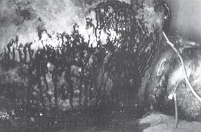
| Fig. 1 - Denuded
bleeding dermis in the posterior region of the thorax (pressure point). |
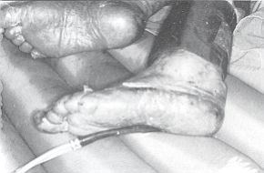
Fig. 2 -
Epidermal necrolysis in both feet. |
|
Previous pathology
On 17 February 1992 the patient had
been admitted to the University Dermatological Clinic for a skin condition from which he
had been suffering for about five years. This condition had recently become intensely
pruritic. The patient was given cortisonic, systemic and local treatment and discharged a
few days later with a diagnosis of mycosis fungoides. The onset of TEN occurred about two
months later.
Clinical course
The clinical parameters monitored were
haematocrit, hourly diuresis, body temperature, arterial pressure and heart rate.
Haernatocrit and hourly diuresis remained normal; body temperature and heart rate were
constantly high; arterial pressure was also constantly above normal, with some critical
episodes requiring immediate pharmacological treatment.
Table I shows the results of the
blood chemistry tests. There was constant hyperglycaernia, together, with in-creased serum
enzymes and bilirubinaemia with values manifesting themselves clinically in the patient's
jaundiced appearance. Plasma creatinaernia values remained normal in spite of the
contemporary increase in urea.
| Date |
4.5.92 |
5.5.92 |
6.5.92 |
7.5.92 |
8.5.92 |
9.5.92 |
10.5.92 |
11.5.92 |
12.5.92 |
13.5.92 |
14.5.92 |
| Urea (mg/dl) |
150 |
70 |
69 |
57 |
104 |
115 |
61 |
106 |
97 |
103 |
95 |
| Glycaemia
(mg/dl) |
138 |
160 |
185 |
192 |
303 |
196 |
211 |
173 |
117 |
145 |
240 |
| Creatinine
(mg/dl) |
1.4 |
0.9 |
0.7 |
0.9 |
1.1 |
0.9 |
1.0 |
0.7 |
0.6 |
0.9 |
0.8 |
| GPT (u/1) |
35 |
29 |
53 |
192 |
179 |
102 |
- |
- |
82 |
129 |
99 |
| GOT. (u/1) |
42 |
35 |
61 |
124 |
113 |
33 |
|
|
49 |
64 |
57 |
| T. bifir.
(mg/dl) |
- |
0.7 |
0.8 |
0.8 |
6.4 |
4.4 |
|
|
3.3 |
4.5 |
3.3 |
| Na+ (meg/1) |
144 |
- |
147 |
138 |
141 |
139 |
139 |
140 |
140 |
144 |
140 |
| K+ (mcgll) |
4.3 |
- |
4.1 |
3.5 |
3.2 |
4 |
3.3 |
4 |
4.8 |
4.1 |
4.4 |
| Plasm. osm.
(mosm/1) |
325 |
310 |
321 |
320 |
322 |
314 |
|
314 |
310 |
319 |
310 |
| Proteins
(g/dl) |
7.5 |
7.2 |
7 |
6.7 |
5.5 |
5.6 |
|
6 |
6.2 |
6 |
4.8 |
| Araylase (u/1) |
|
70 |
60 |
69 |
60 |
55 |
|
- |
88 |
115 |
72 |
|
Table 1
- Blood chemistry |
|
Table II presents the results of the haematological
tests. Note the constant increase in leucocytes, especially in the neutrophil portion, and
the progressive and serious reduction in the number of platelets, with normal red blood
cell and haernoglobin values except on the last date. Lymphocytopenia is present.
| Date |
4.5.92 |
5.5.92 |
6.5.92 |
7.5.92 |
8.5.92 |
9.5.92 |
10.5.92 |
11.5.92 |
12.5.92 |
13.5.92 |
14.5.92 |
| WBC x 10 IlmI |
4.27 |
3.36 |
5.04 |
5.74 |
12.07 |
14.98 |
15.9 |
14.2 |
14.91 |
17.3 |
22.8 |
| RBC x 106/ral |
5.46 |
4.45 |
4.20 |
4.63 |
4.66 |
4.92 |
4.48 |
4.83 |
4.15 |
4.05 |
3.30 |
| Hb (g/dl) |
16.9 |
13.6 |
13.0 |
14 |
14.1 |
14.7 |
14.2 |
14.1 |
12.4 |
11.1 |
10,5 |
| P1t. X W/M1 |
173 |
129 |
114 |
87 |
42 |
26 |
27 |
28 |
42 |
56 |
100 |
| Neut. % |
78 |
82 |
90 |
86 |
90 |
93 |
94.4 |
90,6 |
88 |
90 |
93.1 |
| Lymph. % |
15 |
10 |
5 |
8 |
5 |
5 |
3.2 |
5.4 |
6 |
5.2 |
3.9 |
| Mon. % |
7 |
8 |
5 |
5 |
5 |
2 |
2.4 |
4.0 |
5 |
4.8 |
3.0 |
| Eos. % |
- |
- |
- |
1 |
- |
- |
m |
- |
1 |
- |
- |
|
Table II -
Haematology |
|
In Table III we report the
patient's respiratory condition, which was monitorized by daily blood gas tests. These
showed slight hypoxaemia, with one critical episode resolved by immediate oxygen therapy.
Repeated thorax radiography did not show any important alterations.
| Date |
4.5.92 |
5.592 |
6.5.92 |
7.5.92 |
8.5.92 |
9.5.92 |
10.5.92 |
11.5.92 |
12.5.92 |
13.5.92 |
14.5.92 |
| pH |
7.40 |
7.53 |
7.49 |
7.51 |
7.48 |
7.51 |
7.50 |
7.47 |
7.43 |
7.50 |
7.44 |
| C02 (ERM119) |
24 |
34.6 |
29 |
32.1 |
3. |
39.3 |
38.3 |
37 |
26 |
28 |
28 |
| P02 (inmHg) |
102 |
61 |
64 |
73 |
61 |
52 |
72 |
71 |
65 |
61 |
102 |
| HC03 (MMo'/]) |
14.8 |
29.4 |
21.9 |
26.4 |
26.7 |
25.4 |
30.6 |
26.6 |
23.2 |
21.9 |
19.4 |
| TC02 (mmol/1) |
15.6 |
3 .5 |
22.8 |
27.4 |
21.8 |
30.2 |
31.8 |
27.8 |
24.5 |
22.8 |
20.3 |
| BE (mmoll/1) |
-8.6 |
7.4 |
-0.0 |
- |
-0.7 |
2.0 |
7.7 |
3.2 |
-0.3 |
-0.9 |
-3.8 |
| Sat 02 % |
97.9 |
93.6 |
95.1 |
96.1 |
95.1 |
93.5 |
95.7 |
95.1 |
94.7 |
93.2 |
98.1 |
|
Table III -
Blood gas analysis |
|
Table IV presents the results of
the haemocoagulation tests. Analysis of the Table enables us to evidence a normal
initial haernocoagulative picture, with a hyperfibrinogenaemia compatible with the
clinical condition and without signs of consumption coagulopathy or of hyperfibrinolysis.
The results of tests performed two days later were however compatible with a condition of
consumption coagulopathy with a reduction in the values of prothrombin activity,
fibrinogen, plasminogen, antithrombin III (At 111) and an increase in APTT and FDP.
| Date |
8.5.92 |
10.5.92 |
12.5.92 |
| Prothrombin activity |
75% |
52% |
73% |
| Fibrinogen |
717 rng/dl |
320 mg/dl |
388 rng/dl |
| APTT |
19,3" R.O.82 |
48,5" R.2.16 |
41,8" R.
1.82 |
| PLG |
85% |
60% |
75% |
| At 111 |
68% |
51% |
128% |
| FDP |
8 p g/ml |
40 - 80 pg/ml |
40 - 80 pg/ml |
|
Table IV - Haemocoagulation |
|
The patient initiated heparin treatment
with At III concentrates. Later coagulation tests showed a stabilized picture with
lengthened APTT still present, increased FDP and reduced plasminogen.
Treatment
It was decided to continue antibiotic therapy
(Imipenem instead of the Rolitetracycline previously used) owing to the positivity of
swabs taken from the lesions which in various areas were beginning to show signs of
reepithelialization (Fig. 3) and to the contemporary administration of steroids. Because
of the doubts generated by the previous disease, we began to reduce the daily dosage of
steroids a few days after the patient had been admitted, with a view to their eventual
elimination. Steroids had been administered for a prolonged period and it was not
advisable to interrupt their use suddenly.
TPN was replaced by peripheral nutritional support as the patient was able to feed
himself. This also had the advantage of eliminating the central venous access.
The patient's therapy was completed with ranitidir, vitamins, heparin and At 111, oxygen,
aerosol therapy, bathing and topical treatment.
Haemocultures were performed frequently to detect any sepsis but were all negative, even
if the swabs were positive. This continued until 13 May 1992 when three haemocultures were
positive to Staphylococcus aureus. This positivity was reflected in the
considerable increase in leucocytes (Table II).
Also on 13 May 1992 the patient had a nervous crisis, which according to the consultant
psychiatrist had two possible causes:
- a sense of persecution: the patient having once been
unjustly imprisoned, in the closed atmosphere of the burns centre he was reliving his
previous experience and his loss of freedom;
- steroid psychosis.
The following day, a probable hypertension crisis caused a
cerebral haemorrhage. This haemorrhage, confirmed by a CT scan, was certainly massive,
also considering the altered haemocoagulative picture. The patient was transferred to
Neuroreanimation and submitted to surgery, but died after a few days.
|
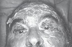
Fig. 3 -
Complete reepithelialization of the face. |
|
Discussion
Aetiology
Piroxicam is a TEN-inducing drug. Out
of 62 cases provoked by non-steroid anti-inflammatory drugs, Roujeau et al. (1) found
piroxicam to be responsible on 13 occasions (20.9%).
Associated lymphoproliferative diseases
As far as we know, mycosis fungoides
has not been observed in association with cases related to TEN, although there have been
various reports concerning lymphoproliferative diseases. An exhaustive bibliography is
given by Bianchi et al. (2), who described a case of TEN in a patient with an anamnesis
that said nothing of drug administration but mentioned a retroperitoneal and pelvic
non-Hodgkin lymphoma discovered on autopsy.
Signs of systemic toxaemia
Fever and tachycardia are part of the
general picture of TEN; this is not true of arterial hypertension and hyperglycaernia,
which in all probability are due to prolonged steroid treatment.
Renal involvement
The high plasma urea levels, with
normal diuresis and normal creatinaemia values, are not compatible with renal dysfunction
and may be related to the condition of hypercatabolism, the acid-base equilibrium
disturbances and the steroid and antibiotic (tetracycline) treatment administered to the
patient.
Revuz et al. (3) believe that the level of plasma urea is an important prognostic factor
because this depends on both renal function and the gravity of stress and catabolism.
Other prognostic factors which, on the basis of the multifactorial analysis that we
carried out, appear to be important are age and the percentage of body surface area
involved.
Renal involvement, when it occurs, is generally secondary to inadequate reintegration of
intravascular volume, which is reduced as a result of the losses caused by the
epidermolytic process. In other words, untreated hypovolaemia causes acute tubular
necrosis with consequent kidney failure.
The rare cases of membranous glomerulonephritis described in the literature (4) and
subsequently cited (5, 6, 7, 8, 9) are not very recent and according to Roujeau et al. (1)
cannot be attributed with absolute certainty to specific TEN-related alterations.
Hepatic involvement
Alterations of transaminase and
bilirubin_ are frequently reported.
In the cases observed by Kvasnicka et al. (10) three patients out of eight presented a
considerable increase in transarninase. Tagami et al. (11) described a case of
ampicillin-induced TEN in which the alteration of hepatic function (increased enzymes and
bilirubin) continued for six months after the patient recovered. The pathogenesis of the
hepatic damage.and, in this particular case, also the pancreatic damage because of the
contemporary increase in amylasaemia, was in the authors' opinion identical to that of the
skin damage and was secondary to the liberation by the patient's lymphocytes, when they
encountered the antigen, of biologically active lymphokines.
All ten patients in the series described by Westly and Wechsler (12) presented a high
level of hepatic enzymes, while five patients out of six - in one of whom the condition
already existed - in the series described by Aub6ck et al. (13) presented altered hepatic
function. In these last cases TEN was induced by allopurinol.
Halebian et al. (14) compared two groups of 15 patients each. One group was treated with
steroids and the other was not. The alteration of the hepatic enzymes was the same in both
groups (nine patients in the treated group and ten in the untreated group). Jaundice
(bilirubin > 5) was clinically evident in one patient in each group. It was
hypothesized that although the mechanism of the alteration was not clear it may have been
due to a primary disturbance rather than to a secondary process.
Saiag et al. (15) found that hepatic enzyme levels were more than doubled in eight out of
fourteen HIV-infected patients suffering from TEN.
Despite numerous reports it is still controversial whether cytolytic hepatopathy (HbsAg
negative) is an integral part of the disease or whether it is not instead directly caused
by a drug, a shock condition or a septicaemic process (9).
Blood and coagulation
The haematological and
haemocoagulative alterations described in single cases and some extensive series also
require further investigtion.
A case of leucopenia associated with thrombocytopenia is reported by Anhalt and Snelling
(16) and also by Fenton and English (17). A case of leucopenia associated with anaemia and
thrombocytopenia is reported by Goens et al. (18) and also by Castelli et al. (19).
Westly and Wechsler (12) observed leucopenia in all ten of their patients. This alteration
occurred on day 2-5 of the disease. Values returned to normal (day 8) in only three
patients, two children and an adolescent not treated with steroids, who recovered from the
disease. Leucopenia was considered to be a cardinal aspect of TEN and its persistence was
regarded as a negative prognostic factor. Age and steroid treatment were also considered
to be related to the outcome.
Lymphopenia, due to a reduction in the number of T lymphocytes, in particular T-helper
lymphocytes, possibly caused by preferential consumption in an immunological conflict, was
described by Roujeau et al. (20) in six cases of TEN.
Bombal et al. (21) carried out a retrospective study of 26 patients suffering from TEN and
found that normochromic and normocytic anaemia and lymphopenia, which may be so marked
that there is a total absence of lymphocytes detectable in the blood, are almost constant
haematological anomalies.
Saiag et al. (15), in their fourteen AIDS patients, found thrombocytopenia (two patients),
neutropenia (two patients) and Jymphopenia (ten patients). Except for lymphopenia these
anomalies cleared up spontaneously.
In short, both the platelets and the red corpuscles or the lymphocytes or the neutrophil
granulocytes, either in or not in association among them, may be altered, mainly in the
sense that they are reduced.
According to Roujeau et al. (9), "anaemia is present in nearly every patient because
of a number of factors, including erythroblastopenia ... lymphopenia is present in 90% of
patients caused by a selective and transitory depletion of the CD4+ T-helper lymphocytes
... thrombopenia in 15%."
Revuz et al. (3) contest the theory that neutropenia has a prognostic significance
independently of other factors.
Halebian et al. (14) found leucopenia with neutropenia in 13 cases. Septicaemia was also
present in 12 cases. It was however pointed out that while on the one hand neutropenia is
important in determining the onset of septic complications it is also true that steroid
treatment may affect the leucocyte count.
Kvasnicka et al. (10) found a normal leucocyte count in two of their eight patients and
leucocytosis in the other six. These researchers however had concentrated their attention
on the patients' haemocoagulative condition, observing signs of disseminated intravascular
coagulation (DIC) of greater or lesser extent in relation to the severity of the clinical
conditions and to the moment when the tests were performed.
Respiratory system
Apart from pneumonia, which is one of the
most frequent complications of TEN, the main alterations of the respiratory system consist
of subclinical interstitial oedema detectable by thorax radiography and mild hypoxia shown
by blood gas test (9).
For this reason Pruitt (6) recommends daily chest Xray and blood gas analysis in order to
monitor the respiratory system so that intubation can be performed immediately if the
necrolytic process begins to affect the mucosa of the iracheobronchial tract.
Conclusion
We should like to stress two main points:
- Toxic epidermal necrolysis is a complex disease: the
alterations in the case we have described (hepatopathy, thrombocytopenia, consumption
coagulopathy) may be independent factors in the disease or they may be related
(hepatopathy and coagulopathy); they may on the other hand be secondary to sepsis which is
frequently a complicating factor in TEN. In our case we found sepsis due to Staphylococcus
aureus, but only after the above alterations had occurred.
- TEN is not usually a disease that receives continuous
treatment. Halebian et al. (14) calculated the time interval between the onset.of the
disease and admission to their burns centre of the two groups of patients they compared
with regard to the use or non-use of steroids. Patients in the first group were admitted
after a mean period of 7.3 ± 0.7 days, and in the second group after 8.3 ± 1.1 days. As
admission to a specialized centre is never immediate, we find ourselves faced with methods
(e.g. the use of central venous access) or therapeutic approaches (antibiotics, steroids)
which may not be advisable. In our patient the steroid treatment was probably responsible
for a condition of psychosis. This would not be the first case, as Halebian et al. (22)
reported that one of the fifteen patients treated with steroids appeared to suffer from
psychosis.
It is also probable that the prolonged steroid treatment
triggered the mechanism which in a situation of coagulopathy not only favoured the onset
of sepsis but also caused the patient to pass from a condition of hypertension to the
acute vascular accident and finally to his death.
RESUME. Les auteurs
décrivent un cas de nécrolyse épidermique toxique (sigle en anglais, TEN) dans un
patient adulte atteint de mycosis fongoïde. Le cours de la maladie, qui touchait 50% de
la surface corporelle et s'est terminé par la mort du patient après un épisode
d'hémorragie cérébrale, était caractérisé par la manifestation d'une grave
hépatopathie, thrombopénie et coagulopathie de consommation. Les auteurs, qui
considèrent ces altérations spécifiques pour la TEN et non secondaires à la condition
septique successivement observée, soulignent la complexité de cette maladie, qui n'est
pas seulement cutanée ou cutanée/muqueuse, et l'importance de l'hospitalisation
immédiate chez un contre spécialisé pour garantir la continuité du traitement
thérapeutique.
BIBLIOGRAPHY
- Roujeau J.C. ', Guillaume J.C., Fabre J.P., Penso
D., Fldchet M.L., Girre J.P.: Toxic epidermal necrolysis (Lyell syndrome). Incidence and
drug etiology in France, 1981-1985. Arch Dermatol., 126: 37 42, 1990.
- Bianchi L., Gatti S., Carrozzo A.M., Marinaro C.,
Iraci S., Luzi F., Nini G.: Sindrome di Lyell e linforna. G. Ital. Dermatol. Venereol.,
125: 255-8, 1990.
- Revuz J., Penso D., Roujeau J.C., Guillaume J.C.,
Rowland Payne C., Wechsler J., Touraine R.: Toxic epidermal necrolysis. Clinical findings
and prognosis factors in 87 patients. Arch. Dermatol., 123:1160-5, 1987.
- Krumlovsky F.A., Del Greco F., Herdson P.B., Lazar
P.: Renal disease associated with toxic epidermal necrolysis (Lyell's disease).Am. J,
Med., 57: 817-25,1974.
- Management of toxic epidermal necrolysis. Lancet
(Editorial), 2:1250-2, 1984.
- Pruitt B.A.: Burn treatment for the unburned. JAMA
(Editorial),257: 2207-8, 1987.
- Ward D.J., Kazeminska E.C., Tanner N.S.B.: Treatment
of toxic epidermal necrolysis and a review of six cases. Bums, 16: 97-104,1990.
- Pousa F., Valero J., Vazquez-Baffo A., Trineado S.:
Bum unit treatment of three Stevens-Johnson syndrome cases with cryopreserved allograft.
Ann. Medit. Burns Club, 5: 160-3, 1992.
- Roujeau J.C., Chosidow 0., Saiag P., Guillaume J.C.:
Toxic epidermal necrolysis (Lyell syndrome). J. Amer. Acad. Dermatol., 23:1039-58, 1990.
- Kvasnicka J., Rezac J., Svejda J., Duchkova H., Kaze
F., Zalud P.,Richter J.: Disseminated intravascular coagulation associated with toxic
epidermal necrolysis (Lyell's syndrome). Br. J. Dermatol.,100: 551-8, 1979.
- Tagami H., Tatsuta K., Iwatski K., Yamada M.:
Delayed hypersen sitivity in ampicillin-induced toxic epidermal necrolysis.
Arch.Dermatol., 119: 910-3, 1983.
- Westly E.D., Wechsler H.L.: Toxic epidermal
necrolysis. Granulocytic leukopenia as a prognostic indicator. Arch. Dermatol., 120:
721-6, 1984.
- Aubbck J., Fritsch P.: Asymptomatic hyperuricaernia
and alloponnot induced toxic epidermal necrolysis. Br. Med. J., 290: 1969-79, 1985.
- Halebian P.H., Corder V., Madden M.R., Finklestein
J.L., Shires G.T.: Improved burn centre survival of patients with toxic epidermal
necrolysis managed without corticosteroids. Ann. Surg,, 204: 50312,1986.
- Saiag P., Caumes E., Chosidow 0., Revuz J., Roujeau
J.C.: Druginduced toxic epidermal necrolysis in patients infected with the human
immunodeficiency virus. J. Am. Acad. Dermatol., 26: 56774, 1992.
- Anhalt G., Snelling C.F.T.: Toxic epidermal
necrolysis. Case Report. Plast Reconstr. Surg., 61: 905-10, 1978.
- Fenton D.A., English J.S.: Toxic epidermal
necrolysis, leucopema and thrombocytopenic purpura - a further complication of
benoxaprofen therapy. Clin. Exper. Dermatol., 7: 277-80, 1982.
- Goens J., Song M., Fondu P., Blum D,, Achten G.:
Haematological disturbances and immune mechanisms in toxic epidermal necolysis. Br. J.
Dermatol., 114: 255-9, 1986.
- Castelli R., Centemeri D., Frattini F., Greco P.:
Quadro mortale in un caso di sindrome di Lyell. Min. Anaest., 52: 345-50, 1986.
- Roujeau J.C., Moritz S., Guillaume J.C., Revuz J.,
Touraine R.: Etude de sous-populations lymphocytaires an cours du syndrome de Lyell. Ann.
Dermatol. Venereol., 110: 185, 1983.
- Bombal C., Roujeau J.C. Kuentz M., Revuz J.,
Touraine R.: Anomalies hématologiques au cours du syndrome de Lyell. Atin. Dermatol.
Venereol., 110: 113-9, 1983.
- Halebian P.H., Corder V., Herndon D.: A burn center
experience with toxic epidermal necrolysis. J. Burn Care Rehabil., 4: 176-83, 1983.
|