Annals ofBurns and Fire Disasters - vol. VIII - n. 4 - December 1995

EXPRESSION OF KERATIN IN POST-BURN SCARS AND KELOIDS

Ramakrishnan M*, Prathiba V.**, Sridhar Rao K.***, Jayaraman V.*, Babu M.**, Gupta P.D.***

* Central Leather Research Institute, Adyar, Madras, India
** Centre for Cellular and Molecular Biology, Hyderabad
*** Child Trust Hospital, Nungambakkam, Madras


SUMMARY. Keratinocyte migration is one of the initial events of re-epithelialization, and therefore an important phase of wound healing. The aim of this study was to consider the expression of keratin, which is the biochemical marker of re-epithelialization. 50 KDa and 58 KDa keratins, which are markers of proliferation, were found to be overproduced in keloids and post-bum hypertrophic scars compared to normal human skin.

Introduction

During epithelialization in wound healing, keratinocytes lateral to the wound margin migrate across the wound bed to cover it. These keratinocytes, which rest on the cutaneous basement membrane zone, migrate upwards to the surface of the skin. During this process they start to differentiate and synthesize keratins. Keratins are marker proteins of all epithelial cells, and are expressed in cell and tissue in a specific manner. They are highly insoluble in most solvents, except high urea concentrations. On the basis of the amino acid sequence, pKI value, etc., keratins can be subdivided into two groups: type 1 keratins, which are 
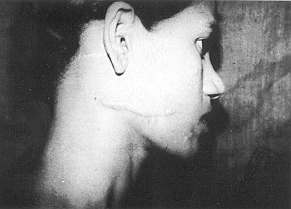
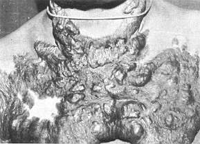
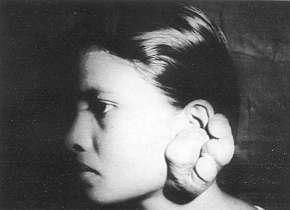
acidic, and type II keratins, which are basic.
Basal cells express K5 (58 KDa) and K14 (50 KDa),' a pair of acidic and basic proteins which are markers for proliferation. Type II keratin KI (67 KDa) and type 1 keratin KIO (56.5 KDa) are expressed in abundance only in terminally differentiating epidermal cells. The present study deals with the expression of keratins in post-burn hypertrophic scars and keloids.

Materials and methods

Clinical specimens

The tissue samples were obtained from Child Trust Hospital, Madras, and informed consent was obtained from each patient. All the patients providing tissue for this study were of Indian origin. The lesions were diagnosed as keloids or post-burn scar on the basis of clinical appearance and also by the over-expresSiOn of fibronectin (Table I, Fig. 1).

Keratin extraction. SDS-Page and Western analysis

A number of 6 mm punch biopsy specimens were obtained from the tissue and left in PBS at 60 'C for 3-5 rninutes. The epidermis was peeled off and extracted once with high salt detergent solution, sonicated briefly and kept for overnight extraction. The pellet obtained on centrifugation was washed with low salt buffer. After washing, keratin was extracted by boiling the pellet in IX sample buffer containing 8M urea for 10 minutes. The extracts were subjected to SDS-PAGE1 and transferred to nitrocellulose.' Incubation of the nitrocellulose blot with polyclonal rabbit keratin antiserum was carried out with 1 in 200 dilutions in PBS solution containing 2.5% BSA at room temperature for one hour. After extensive washing with PBS containing 0.2% Tween, bound antibodies were detected by incubation with radio-iodinated protein A. Finally the nitrocellulose blot was washed extensively, dried and subjected to autoradiography.

RNA extraction and hybridization

Total RNA was extracted from the epidermis according to the procedure of Choinczyriski et al.' For slot-blot hybridization the RNA samples were applied onto nylon membranes. The blots were then prehybridized and hybridized with keratin cDNA. After washing, the blot was autoradiographed at -70 'C using X-ray films and intensifying screens.

N'

Patient age (yr)

Sex

Biopsy Site

Months after scar

1

22

F

Thigh

-

2

30

M

Abdomen

-

3

18

F

Abdomen

-

4

25

F

Face

24

5

35

F

Face and neck

30

6

24

M

Hand

17

7

18

F

Earlobe

60

Table I - Source of human normal skin, post-burn hypertrophic scar and keloid tissues


Fig. la - Post-bum hypertrophic scar Fig. lb - Post-bum hypertrophic scar
Fig. lc - Keloid Fig. 1d - Keloid


Fig. 2 - SDS-PAGE of keratins isolated from epidermis of normal (lanes 2, 3, 8), HTS (4, 5, 6) and keloid (lane 7) Marker proteins (lane 1). (a) phosphorylase-13 (97,400); (b) bovine serum albumin (67,000); (c) ovalburnin (43,000); (d) carbonic anhydrase (30,000); (e) trypsin inhibitor (20,000). Fig. 2 - SDS-PAGE of keratins isolated from epidermis of normal (lanes 2, 3, 8), HTS (4, 5, 6) and keloid (lane 7) Marker proteins (lane 1). (a) phosphorylase-13 (97,400); (b) bovine serum albumin (67,000); (c) ovalburnin (43,000); (d) carbonic anhydrase (30,000); (e) trypsin inhibitor (20,000).

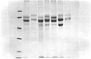
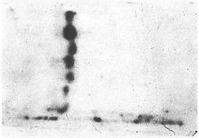
Results

Fig. 2 shows the SDS-PAGE analysis of keratin extracted from normal skin, post-burn hypertrophic scar (HTS) and keloids, as described in Materials and Methods. The polypeptide bands in all the tissues are seen spread in the 50-68 KDa range. There is no difference in the number of polypeptide bands in all the tissues, but it was observed in Coomassie Blue stained preparations that bands with molecular weight 58 and 50 KDa were stained densely in post-burn scar (lanes 4, 5, 6) and keloid (lane 7) when compared to normal skin (lanes 2, 3, 8).

In order to cheek whether these polypeptides in SDS gels belong to keratin protein, we used polyclonal antibodies against human keratins to observe the cross-reactivity of these antibodies with the polypeptide bands. Fig. 3 shows the cross-reactivity of human antikeratin antibodies with the keratin extracted from all three different types of tissues. The 58 KDa shows thick bands in Western-blot analysis in post-burn scar and keloid tissues.

Fig. 3 - Immuno blot of keratins in normal (lanes 1, 2, 7), HTS (lanes 3, 4, 5) and keloid (lane 6). Primary antibody used was polyclonal rabbit anti-human keratin anti-body. Secondary antibody was `1 labelled protein A. Fig. 3 - Immuno blot of keratins in normal (lanes 1, 2, 7), HTS (lanes 3, 4, 5) and keloid (lane 6). Primary antibody used was polyclonal rabbit anti-human keratin anti-body. Secondary antibody was `1 labelled protein A.
Fig. 4 - Slot-blot analysis of keratin RNA expression in normal, hypertrophic scar and keloid epidermis. Total mRNA was extracted from normal (lane 1), HTS (lane 3 ) and keloid (lanes 2 and 4) by the GITC/phenol-chloroform extraction method. A serial dilution of RNA for each sample was applied directly onto a nylon membrane. The blot was hybridized with DNA specific for 50 kDa keratin (PKB-2) 1425 bp cloned into pst- I site on pl3R 322. Fig. 4 - Slot-blot analysis of keratin RNA expression in normal, hypertrophic scar and keloid epidermis. Total mRNA was extracted from normal (lane 1), HTS (lane 3 ) and keloid (lanes 2 and 4) by the GITC/phenol-chloroform extraction method. A serial dilution of RNA for each sample was applied directly onto a nylon membrane. The blot was hybridized with DNA specific for 50 kDa keratin (PKB-2) 1425 bp cloned into pst- I site on pl3R 322.

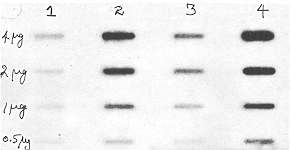
For further confirmation, RNA extracted from all the tissues was hybridized with cDNA for 50 KDa keratin. Slot-blot analysis (Fig. 4) showed that mRNA keratin is overexpressed in post-burn scar and keloid compared to normal (lanes 2 and 4 versus lanes 1 and 3).

Discussion

Re-epithelialization is an important event during wound healing and is accomplished as epidermal cells migrate over the provisional matrix provided by granulation tissue and re-establish the basement membrane.
The basal keratinocytes initially avoid the differentiation and migrate laterally to cover the wound. In keratinocyte proliferation and differentiation, the undifferentiated proliferating stem cells are closer to the supporting dermal layer, while the cells that have migrated away from the basal layer become differentiated and cease to proliferate.
Our present study indicates that in abnormal wound healing the expression of keratin is enhanced. In keloid and post-bum hypertrophic scar 58 KDa keratins are found to be overproduced. This was confirmed by the Westernblot analysis. Slot-blot analysis shows that the message for the expression of 50 KDa is more at the RNA level, which implies that there are more undifferentiated keratinocytes in post-bum hypertrophic scar and keloid. Since epithelial cells are intimately associated with connective tissue elements in vivo, it is possible that epithelial cell-extra cellular matrix interactions play an important role in determining differential keratin expression.
Detailed study of the expression of basement membrane components such as collagen IV and laminin is in progress.


RESUME. La migration des kératinocytes est un des premiers événements de la ré-épithélialisation et pour cette raison c'est une phase importante de la guérison des lésions. Les auteurs, dans cette étude, ont voulu considérer l'expression de la kératine, qui est le marqueur biochimique de la ré-épithélialisation. Ils ont trouvé qu'il y avait une surproduction des kératines 50 KDa et 58 KDa, qui sont les marqueurs de la prolifération, dans les chéloïdes et les cicatrices hypertrophiques dues aux brûlures en comparaison de la peau humaine normale.


BIBLIOGRAPHY

  1. Stenn K.S., Depalma L.: Re-epithelialisation. In: "The Molecular and Cellular Biology of Wound Repair" (Clark R.A.F., Hensen P.M., Eds), pp. 321-35. Plenum Press, New York, 1988.
  2. Clark R.A.F.: Cutaneous tissue repair: Basic biologic considerations. J. Am. Acad. Dermatol., 13: 701-25, 1985.
  3. Uitto J., Oikarinen A., Thody AJ.: Scientific Basis of Dermatology: A Physiological Approach (Thody A.J., Friedmann P.S., Eds), Churchill Livingstone (date not supplied).
  4. Fuchs E.V., Coppock S.M., Green H., Cleveland DW.: Two distinct classes of keratin genes and their evolutionary significance. Cell, 27: 75-84, 1981.
  5. Hamikoglu LE, Fuchs E: The cDNA sequence of a type 11 cytoskeletal keratin reveals constant and variable structural domains among keratins. Cell, 33: 915-24, 1983.
  6. Nelson W., Sun T.T.: The 50 and 58-kDalton classes as molecular markers for stratified squamous epithelial cell culture studies. J. Cell Biol., 97: 244-51, 1983.
  7. Laerninli U.K.: Cleavage of structural proteins during the assembly of the head of bacteriophage T4. Nature, 76: 4350-4, 1979.
  8. Towbin H., Staclein T., Gordon J.: Electrophoretic transfer of proteins from acrylamide gels to nitrocellulose sheets, Procedure and applications. Proc. Natl. Acad. Sci. USA, 1979.
  9. Chomczynski P., Sacchi N.: Single step method of RNA isolation by acid guandinium thiocyanate -phenol -chloroform extraction. Anal. Biochem., 162: 156-9, 1987.
  10. Fuchs E., Albers K., Kopan R.: Terminal differentiation in cultured human epidermal cells. Adv. Cell Cult., 6: 1-33, 1988.
  11. Kurpakus M.A., Stock E.I., Jories J.C.R.: The role of basement membrane in differential expression of keratin proteins in epithelial cells. Dev. Biol., 50: 243-55, 1992.
This paper was received on 14 February 1994.

Address correspondence to: M. Ramakri~shnan M.D., Central Leather Institute, Adyar, Madras, India.


THE THIRD PAN ARAB CONGRESS OF PLASTIC AND RECONSTRUCTIVE SURGERY AND BURNS
21 - 24 March 1996 - Beirut

Main Topics:
Aesthetic surgery
Maxillofacial and head and neck surgery
Reconstructive surgery and ruicrosurgery
Hand surgery Burns

Scientific secretariat: B. Atiyeh M.D., c/o AUB New York Office,
850 Third Avenue, 18th Floor, New York, NY, 10022 USA
Fax: 1 (212) 478 1995


Contact Us
mbcpa@medbc.com