| Annals of Burns and Fire Disasters - vol. IX - n. 1 - March 1996
MANAGEMENT OF SEVERELY BURNED PATIENTS A STUDY OF 684
SEVERELY BURNED PATIENTS ADMITTED IN THE LAST SIX YEARS TO THE BURN AND PLASTIC SURGERY
CENTRE, TRIPOLI, LIBYA
Zaidi M.M., Abussetta A.A., Franka MR, Shahata G,
Traikov E, Uyang L.
Burn and Plastic Surgery Centre, Tripoli, Libya
SUMMARY. Between 1 January 1989
and 31 December 1994 684 severely burned patients were admitted to the Intensive Care Unit
(ICU) in the Burn and Plastic Surgery Centre in Tripoli (Libya). The ICU is an isolated
department with nine beds (three single isolated beds, plus six others), with separate
operating theatres and five air-fluidized beds. The Unit is staffed by four
anaesthesiologists from the Anaesthesia Department together with three burn surgeons
working exclusively in the Unit, as well as specially trained nurses. All facilities for
non-invasive monitoring are available for the nine beds. As in the central control unit,
dialysis is available in the ICU if required.
Criteria for admission to the Intensive
Care Unit
- Severe burns in more than 30% TBSA
- Bums in extremes of ages
- Bums with smoke inhalation
- High-voltage electric burns
- Bums associated with other medical problems
Materials and methods
In the 6-year period between 1 January
1989 and 31 December 1994, 684 patients, including 249 with smoke inhalation, were
admitted to our ICU. All patients were managed by the Evans formula, modified in children,
as follows:
- Ringer's lactate 1 ml/kg/% TBSA/24 h
- Plasma protein 1 ml/kg/% TBSA/24 h
- Dextrose saline according to body weight
- (half of this amount is given in the first 8 hours)
All patients with smoke inhalation or
suspected smoke inhalation are either immediately intubated or prepared for intubation.
All patients are bathed in the emergency room after stabilization and before admission to
the ICU. Escharotomy is also decided if there is a deep burn around the extremities or the
chest wall. Analgesia and sedation are usually initiated in the emergency room.
In most cases wound management is performed by early excision 12 hours post-burn.
Surgery is completed in the first nine days post-burn in patients with a moderate
percentage of burned body surface area. Temporary wound closure is performed with homoskin
graft (usually from volunteer donors), xenograft (prepared from sheep), or synthetic skin
substitute (Biobrane). Nutrition is initiated after 12 hours either orally or by
nasogastric tube with parenteral caloric support. We analyse our patients according to age
(Table I), sex (Table II), percentage burn (Table III), cause of burn
(Table IV% presence of smoke inhalation and related mortality (Table V~, and
the mortality rate for patients admit ted to the ICU (Tables VI, VII). We also
analyse the direct cause of death in each group and describe our methods of dealing with
smoke inhalation.Smoke inhalation is suspected in cases of flame burn in closed spaces,
burned face, soot in the nostrils or sputum, stridor, hoarseness or loss of voice, and
respiratory insufficiency. We diagnose smoke inhalation by:
- history (fire in closed spaces)
- physical findings (facial burns, ocular irritation, sooty
sputum, stridor and respiratory insufficiency)
- laboratory investigations (arterial blood gases, carbon
monoxide rate)
- pulmonary function tests (reduction of forced expiratory
volume)
- fiberoptic bronchoscopy (showing level of injury)
We achieve better oxygenation in patients
suffering from smoke inhalation by increasing P02 and improving perfitsion ventilation.
Systemic toxication is treated as shown below.
| Gas |
Source |
Effects |
Carbon monoxide
Carbon dioxide |
Any organic matter |
Tissue hypoxia
Narcosis |
Hydrogen chloride
Hydrogen cyanide |
Plastics
Wool, silk, nylon |
Mucosal irritation
Respiratory failure
Coma |
| Benzene |
Petroleum, plastics |
Mucosal irritation
Coma |
| Ammonia |
Nylon |
Mucosal irritation |
|
Toxic elements in
smoke |
|
Initial treatment consists of an
aggressive approach to upper airway maintenance and pulmonary support by:
- nasal endotracheal tube for bronchial lavage and adequate
oxygenation
- connection to ventilator with 5 cm water PEEP to maintain
airway patency and adequate functional residual capacity
- bronchodilators such as sulbotamol (Ventoline) via aerosol
- Lasix to minimize pulmonary oedema
- antibiotics to prevent secondary infection
- Ca channel blockers (e.g. Verapamil) to improve the
vascularity of pulmonary and tracheal membranes
Results
Table I shows that most of the
patients admitted to the ICU in the six-year period were over 14 years of age. There were
however also paediatric patients - we admitted 18 infants aged less than one year, mainly
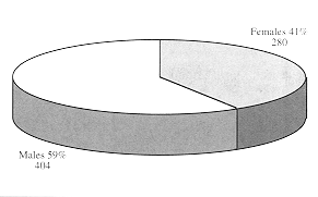
suffering from scald burns. The majority of the patients were males, owing to the fact
that most of the cases were related either to flame burns or high-voltage electric burns
caused by accidents at work (Fig. 1, 2, 3, 4).
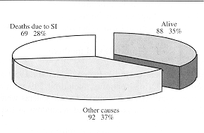
Out of all patients admitted to the ICU, 249 complained of smoke inhalation, requiting
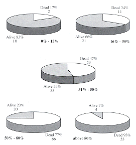
special management. The mortality rate was about 40% of the whole population, and was
related to the percentage of TBSA burned. In the entire series, only five patients out of
54 with TBSA percentage over 80% survived, and three of these presented smoke inhalation.
 |
 |
| Fig. 1
- Mortality rate in relation to TBSA burned |
Fig. 2 -
Causes of death. |
 |
 |
| Fig. 3 -
Overall distribution of patients by sex. |
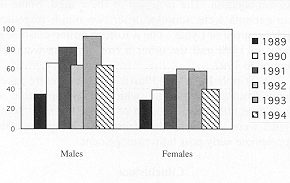
Fig. 4
- Distribution of patients by sex by year. |
|
| Year 1 % TBSA |
0-15 |
16-30 |
31-50 |
51-80 |
81-100 |
| 1989 |
2 |
18 |
20 |
15 |
9 |
| 1990 |
6 |
27 |
28 |
29 |
15 |
| 1991 |
11 |
53 |
39 |
26 |
7 |
| 1992 |
11 |
37 |
44 |
25 |
7 |
| 1993 |
15 |
35 |
56 |
32 |
13 |
| 1994 |
10 |
25 |
32 |
30 |
7 |
| Total |
55 |
195 |
219 |
157 |
- |
|
Table I
- Intensive care unit burn patients - distribution by age |
|
The mortality rate was low when the
percentage TBSA burned was less than 50%. In patients with TBSA burned between 51% and
80%, there was a major improvement in the mortality rate in the six-year period (Table VII).
Not many patients died in the resuscitation phase, 4 most of the deaths occurring after
the second week secondary to septicaernia, multiorgan failure, and bleeding from the
gastrointestinal tract. Two of the deceased patients were pregnant, which suggests that
severe burns associated with pregnancy result in high mortality (we intend to study this
phenomenon in further studies). In the six-year period, our experience was that bleeding
from the gastrointestinal tract was a real problem. We lost five patients as a result of
gastrointestinal bleeding. These were not possible candidates for laparotomy, be cause of
their general condition. As a routine measure we treat all our patients prophylactically
against bleeding.
As shown in Table V and VI, 53 out of 54 patients with more than 81% TBSA burns
also complained of smoke inhalation. The majority of these died. Some of these patients
were admitted after two major tragedies which occurred in Lybia - one a Korean airline
crash near Tripoli in 1990 and the other a fire during a wedding reception in 1992. The
Tripoli Burn and Plastic Surgery Centre takes patients from all over Lybia, which has a
population of around five millions in an area of 2,000,000 kin'. Some patients were
transferred late from other hospitals after inappropriate early post-burn management.
| Year / Causes |
Flame |
Scald |
Electric |
| 1989 |
50 |
14 |
- |
| 1990 |
84 |
19 |
2 |
| 1991 |
92 |
42 |
2 |
| 1992 |
91 |
33 |
- |
| 1993 |
117 |
23 |
11 |
| 1994 |
73 |
23 |
8 |
| Total |
507 |
154 |
|
|
Table II -
Intensive care unit burn patients - distribution by sex |
|
| Year / Age (yr) |
0-1 |
1-3 |
3-6 |
6-14 |
14 > |
| 1989 |
1 |
11 |
9 |
10 |
33 |
| 1990 |
1 |
11 |
11 |
14 |
68 |
| 1991 |
5 |
20 |
26 |
13 |
72 |
| 1992 |
4 |
28 |
8 |
14 |
75 |
| 1993 |
3 |
18 |
18 |
17 |
95 |
| 1994 |
4 |
16 |
9 |
11 |
64 |
| Total |
18 |
99 |
81 |
79 |
- |
|
Table III
- Intensive care unit burn patients - distribution by percentage TBSA burned |
|
| % TBSA |
Age
(yr) |
0-1 |
1-3 |
3-6 |
6- 14 |
14 |
| 0-15 |
Surv. |
1 |
- |
1 |
2 |
6 |
| |
Dec. |
- |
- |
- |
1 |
1 |
| 16-30 |
Surv. |
|
4 |
3 |
8 |
6 |
| |
Dec. |
|
3 |
2 |
1 |
5 |
| 31-50 |
Surv. |
|
2 |
2 |
4 |
25 |
| |
Dec. |
1 |
4 |
7 |
2 |
15 |
| 51-80 |
Surv. |
- |
- |
1 |
3 |
16 |
| |
Dec. |
1 |
6 |
8 |
8 |
43 |
| 81-100 |
Surv. |
- |
- |
- |
- |
4 |
| |
Dec. |
- |
- |
1 |
3 |
49 |
| Total |
|
3 |
19 |
25 |
32 |
170 |
| Surv. |
|
1 |
6 |
7 |
17 |
57 |
| Dec. |
|
2 |
13 |
18 |
15 |
113 |
| Total number surv. =
88 |
| Total number dec. =
161 |
| Total number = 249
(Surv. survived Dec. deceased) |
|
Table IV -
Intensive care unit burn patients - distribution by cause of burn |
|
Year |
Male |
Female |
1989 |
35 |
29 |
1990 |
66 |
39 |
1991 |
82 |
54 |
1992 |
64 |
60 |
1993 |
93 |
58 |
1994 |
64 |
40 |
1 |
404 |
280 |
|
Table V
- Mortality rate related to smoke inhalation |
|
| % TBSA |
Age
(yr) |
0-1 |
1-3 |
3-6 |
6-14 |
14 > |
| 0-15 |
Surv.
Dec. |
6
- |
15
1 |
5
1 |
4
3 |
15
5 |
| 16-30 |
Surv.
Dec. |
6
3 |
38
11 |
28
11 |
22
2 |
57
17 |
| 31-50 |
Surv.
Dec. |
-
1 |
18
9 |
10
12 |
15
8 |
102
44 |
| 51-80 |
Surv
Dec. |
-
2 |
-
7 |
2
11 |
10
12 |
39
74 |
| 81-100 |
Surv.
Dec. |
-
- |
-
- |
-
1 |
-
3 |
5
49 |
| Total |
|
18 |
99 |
81 |
79 |
407 |
| Surv. |
|
12 |
71 |
45 |
51 |
218 |
| Dec. |
|
6 |
28 |
36 |
28 |
189 |
Total number sury. =
397
Total number dec. = 287
Total number = 684 (Surv. = survived Dec. = deceased) |
|
Table VI -
Overall mortality rate |
|
| Year 1 Outcome |
Survived |
Deceased |
| 1989 |
38 |
26 |
| 1990 |
51 |
54 |
| 1991 |
71 |
65 |
| 1992 |
68 |
56 |
| 1993 |
95 |
56 |
| 1994 |
66 |
38 |
| Total |
389 |
295 |
|
Table VII
- Outcome of management of ICU patients |
|
Conclusions
The incidence of severe burns is
increasing in thirdworld countries as a result of economic progress. The number of cases
and the severity of burns are increasing, and this requires a national programme of
prevention and management. Regional co-operation in this field is also essential.
Mortality could be decreased by
improving burn services and developing specialized burn units. Our experience is that the
treatment of burns in general surgery departments fails to provide the necessary
facilities, while some physicians tend to prefer straightforward general surgery to the
complex management of difficult burns. We feel very strongly that proper resuscitation in
the early postburn phase is decisive in the outcome of treatment. Many patients die
because of improper management during the resuscitation phase, and this requires education
of the medical personnel in general hospitals with regard to burn problems, clear
management plans, charts, guidelines to the transfer of patients, etc. Emergency
departments must be in a condition to provide all this. Early transfer to burn units of
patients in better condition will reduce the morta~ lity rate.
It was proved that early surgical
management of the wound considerably improved our results.
Early nutritional support (oral with
parental nutrition) had a major effect on our results and we therefore consider it
essential.
RESUME. Dans la période 1
janvier 1989 - 31 décembre 1994 684 grands brûlés ont été admis au
service de réanimation du Centre de Brûlés et de Chirurgie Plastique à Tripoli
(Libye). Le service de réanimation est un secteur isolé avec neuf lits (trois lits seuls
isolés, plus autres six), avec ses salles d'opération et cinq lits à air fluidisé. Le
personnel du service de réanimation se compose de quatre anesthésiologistes du Service
d'Anesthésie, trois chirurgiens spécialistes des brûlures qui travaillent exclusivement
pour le service de réanimation, et des infirmiers spécialisés. Toutes les opérations
du monitorage non-invasif sont disponibles pour tous les neuf lits. Au besoin la dialyse
peut être pratiquée dans le service, comme dans l'unité centrale de contrôle.
BIBLIOGRAPHY
- Clark W.R.: Smoke inhalation diagnosis and treatment. World
J. Surg., 16: 22-9, 1992.
- Clark W.R., Bonaventura M., Myers W.: Smoke inhalation
management and airway management at a regional burn unit. J. Burn Care Rehabil., 11:
121-34, 1990.
- Hill I.R.: Particulate matter of smoke inhalation. Ann.
Acad. Med.Singapore, 22: 119-23, 1993.
- Joannovich J., Kastana D., Alexakis D., Tsoutsos D.,
Panayotou P.: Pregnancy and burns: experience from five cases. Ann. Medit. Burns Club, 7:
141-2, 1994.
- Lalonde C., Knox J., Demling R.: Burn edema is accentuated
by moderate smoke inhalation in sheep. Surgery, 112: 908-17, 1992.
- Morris S.E., Navoratnom N.: Comparison of the effect of the
burn injury and smoke inhalation on bacterial translocation. J. Trauma, 30: 639-43, 1992.
- Napoli B., D'Arpa N., Gullo S., Masellis M.: Epidemiology,
clinical treatment and therapy in electrically burned children. Ann. Medit. Burns Club, 7:
188-93, 1994.
- Peitzman A.B., Shires G.T.: Smoke inhalation evaluation of
radiological manifestation and pulmonary dysfunction. J. Trauma, 291232-8, 1989.
- Ruddy R.M.: Smoke inhalation injury. Pediat. Clin. North
Am.:317-36, April 1994.
- Zaidi M., Abussetta D.R., Brogowski A., Franka MR.:
Analysis of burned children treated in the Bums and Plastic Surgery Centre, Tripoli,
Libya, in the year 1992, Ann. Medit. Burns Club, 6: 217-23, 1993.
| This paper was received on 4 May
1995. Address correspondence to: Dr
Mustafa Zaidi
Head of Department, Bum and Plastic Surgery Center
Tripoli, Libya. |
|