| Annals of Burns and Fire Disasters - vol. IX - n. 2 - June 1996
EXPERIMENTAL BURN MODELS
Santos Heredero FX, Hamann C., Obispo Martin
J.M., Rodriguez Arias C., Coca Menchero S.
Experimental Surgery Unit, Hospital Universitario del
Aire, Madrid, Spain
SUMMARY. The
experimental study of burns requires the use of different animal models in order to
reproduce physiopathological conditions. The basic animal models and their manipulation
are presented based on the experience of our Experimental Unit. The most frequently used
animal is the Wistar rat due to its availability, low cost, resistance to infections, and
the feasibility ofreproducing different types of burns. Scalding is the easiest mechanism of
provoking a dermal experimental burn. The possibility of varying water temperature,
time of exposure and the burned area makes this method ideal for reproducing almost every
kind of thermal aggression. Electrical burns usually require superior animals to achieve
lesions comparable to those observed in humans. To evaluate the depth of the experimental
burn, the Suzuki scale is used, which gives objective histological parameters of the
lesion. With this scale, the scalding burn model most frequently used in our Unit provokes
superficial or deep dermal lesions depending on exposure time and temperature.
Catheterization of the common femoral vein has proved to be the most viable way of
obtaining blood samples, providing a long-term route for blood drawing or drug
administration. If the experimental burn models described are to be homologated and
considered comparable, they inusi be reproducible by other researchers, consistent in
results, simple in performance, and, if possible, low in cost. This last condition is
easily achieved with the models described, and makes it possible to extend animal
experimentation in the field of burns to units and centres which are not specialized and
have only limited resources.
Introduction
The objectives of animal
experimentation in the field of burns research have changed considerably in the past
decades. In the 1960s and 1970s the main interest of most studies centred around
physiopathology and in particular the treatment of post-burn hypovolaemic shock.',' There
now exist effective fluid resuscitation methods that make death an exception in the first
stages of hypovolaemic shock. Present-day investigators have directed their efforts
towards the analysis of the mediators - mainly chemical -of the systemic response which
occurs after thermal aggression. These mediators, first descibed by Ninneman,' have been
shown to play an essential role both in immune alterations and in post-burn metabolic or
vascular modifications. For this reason, the majority of current lines of investigation
are directed towards identifying these mediators and evaluating their role in the
different alterations that occur in the immediate or late stages of the burn patient's
general illness.
Once the initial resuscitation phase is overcome, infection is today the primary cause of
death in bum patients. For this reason, now as in past decades, the study of infection
constitutes the second largest field of investigation. It should be noted that the
chemical mediators, as modulators of the immune response, also play an essential role in
these studies.
As can be seen, burns are still a very large field for research. This research often
requires the use of animals, for which reason the development of reliable experimental
models is an absolute requirement.
The objective of any experimental model is the reproduction of the physiopathological
conditions of a burn, with the aim of analysing its local or general repercussions, and,
where possible, of modifying them.
Animal models
The rat is the most widely used animal
in the experimental study of physiopathological or therapeutic attitude situations. The
experimental rat is the Rattus norvegicus, of the order Rodentia, family Muridae, genus
Rattus. The most frequently used strains are Wistar and SpragueDawley. The Wistar rat is
an albino, with a thick head and a tail that is shorter than the body; it is very
resistant to infection, which makes it the ideal specimen for the study of burns,
especially when prolonged survival is required. The Sprague-Dawley rat, also an albino,
with a fine head and a long tail, is on the contrary very susceptible to infection,
especially ofthe respiratory (ract.
The rabbit, Oryctolagus cuniculus, order Lagoniorpha, family Leporidae, genus Oryclolagus,
presents more than 70 varieties for use in laboratories,, from the giant Flanders rabbit,
weighing more than 5 ko, to the minute Dutch rabbit. Experimental rabbits are usually
albino, with rounded body, elongated head, long ears and short (ail, and longer hindlegs
than forelegs. These rabbits have a high risk of infection, and they therefore require
much more care in their manipulation than rats. Surgical manipulation in rabbits must take
place in sterile conditions, a requirement not always essential in rats. However, the
price of rabbits is approximately ten times higher than that of rats, and this greatly
increases the difficulty of using rabbits in research units with limited financial
resources.
Pigs are being used with increased frequency as experimental animals, especially such
varieties as the Mini Pig, due to its great anatomical and physiological similarity to
human beings. In the study of bums, we consider that the combination of their high cost,
anaesthetic requirements and post-operative care does not make them ideal research
animals, especially in units not possessing important infrastructures. In our unit, pigs
and rabbits have been used as experimental models for the production of ear burns,
according to Kaweski's technique,' and for the evaluation of the penetrating capacity of
various antimicrobial agents.'
Anaesthesia
Although anaesthesia introduces a
series of factors and variables which theoretically could modify experimental results, it
is compulsory in animal burn models, in accordance with the requirements of Directive
86/609 of the Council of European Communities.
The rat is easily anaesthetized by inhalation of ether or methoxylfluorane. Both these
agents are effective, but the use of ether in particular occasionally makes the prolonged
maintenance of anaesthesia difficult; they both also present a high risk of causing
ventilatory insufficiency with an important increase of tracheobronchial secretion. In our
experience, intraperitoneal anaesthesia is the most advisable, as it allows good control
of sedation and analgesia levels with few cardiorespiratory depression problems. The
solution used consists of 25 ing Ketamine, 4 mg Diazepam, and 1 mg Atropine, at a dose of
0.2 ral per 100 g body weight. This dosage may be supplemented with successive fractions
to maintain prolonged periods of anaesthesia. In rabbits we use sodium pentobarbital at a
dose of 30 mg per kg of body weight by intraperitoneal or intravenous route through the
dorsal vein of the ear.
Experimental burn models
In our unit we have used both thermal
and experimental burn models for the study of local and systemic response to the
aggression.
The thermal model that we prefer is scalding. In our opinion this model offers conditions
of facility, reliability and control that make it superior to all others. We produce the
thermal lesion on the back of the animal as follows: after anaesthetization of the animal
an electric shaver is used to expose a cutaneous surface on the back, which constitutes
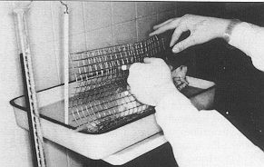
30% of TBSA; the rat is then placed on its back in a mouldable metal wire cage (Fig. 1).
This device is a modification of that described by Walker.' Once the animal is securely
immobilized in the metal cage, the shaved dorsal area is submerged for 12 see in water at
70 'C. This model inflicts a deep dermal burn in the entire cutaneous area exposed.
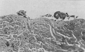
Histologically, skin burned in this way shows the classic signs of epidermic necrosis, a
diffuse perivascular infiltrate, and an important level of collagen degeneration at the
papillary dermis (Fig. 2). The model allows variation in exposure time and water
temperature in order to obtain thermal lesion of different depths.
 |
 |
| Fig.1 - Metallic cage
for the submersion of the animal in hot water. |
Fig.2 - Histology of skin burned by
scalding with water at 70 °C for 12 seconds showing epidermal necrosis, diffuse
perivascular infiltrate and important collagen degeneration at the level of the papillary
dermis, characteristic of a deep dermal burn. |
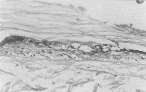 |
Fig.3 - Histology of skin burned by
scalding with water at 70 °C for 6 seconds. |
|
This type of thermal lesion causes au
inilial pliase of' massive fluid extravasation, which leids to the deatli ofilic animal
due to hypovolaemic shock iii the first 6-9 hours unless lost fluids are replaced. If a
more prolonged followup is required, it is necessary to install a fluid resuscitation
regime, for which we administer 1.0 itil of 0.9% NaCI intraperitoncally immediately
post-burn. The second method of thermal burn used in our Unit produces the lesion by means
of direct contact. We use the model described by Feifel.1 The back of the animal is
shaved, and a copper disk (diameter 4 cm), heated to 250 'C, is applied to the skin as
many times as necessary to burn the desired surface area. The major drawback of this
method is the difficulty of ensuring a constant pressure of the disk on the skin in
successive applications, so that it is almost impossible to obtain a burn of uniform
depth.
In 1991 Suzuki et at.' described a burn method based on skin contact with a glass chamber
through which water circulates at a predetermined temperature. This allows application at
a constant pressure of 10 g/CM2. The great advantage of this model is the possibility of
varying temperature and exposure time, as required by the researcher, and also of applying
higher or lower contact pressure, which makes it possible to add ischaerma to the thermal
lesion. This model allows evaluation of the lesion component due to temperature,
dissociating it from that due to compression ischaernia.
The experimental study of chemical burns in the past presented the inconvenience of the
uncontrollable diffusion of the chemical agent beyond the area to which it was applied.
This prevented the availability of a contrastable and uniform model. However, in 1994 Kim
et at."' described a model for the experimental analysis of the effects of chemical
agents on the skin that solved this problem. This model consists of a Teflon hernicylinder
with a window at the cemre. The rat is placed on the hernicylinder and the window is made
to coincide with the area of shaved skin on which the chemical agent is administered. A
pressure cuff is placed on the ventral side of the animal, with the aim of firmly adhering
the back to the Teflon hemicylinder, thus preventing the chemical agent from escaping
beyond the limits of the open window. The area of the window is calculated as a function
of the surface area that the researcher wishes to expose to the chemical agent, on the
basis of the following formula:
Aperture area = (total BSA x % BSA injury desired)/100
With this model it is possible to test different chemical
aggressors and, by varying the exposed skin surface, to evaluate local and systemic
responses.
The study of electric burns generally requires superior animals in order to allow
application of currents with similar effects to those suffered by humans. In 1990 Chilbert
et al.` introduced the application of electrical currents of 1 A intensity at 60 Hz in the
legs of dogs. This model shows a 70% reduction in muscle impedance, which suggests an
irreversible necrosis of myocytes. Possibly the most complete study of the effects of the
experimental application of electric Current Wils published in 1988 by Zelt et a].
12 These authors apply electric currents (40 0, 3500 V and 4.2 A) for 2.5 see to the upper
extremities of primates. The most interesting part of their work is their exhaustive
analysis of' the effects of' these currents. They first performed a histological analysis
of the skin and muscle, which revealed patchy cellular necrosis intermixed with viable
cells. At the same time they performed a digital subtraction angiography, demon strating
segmental narrowing and "pruning" of large vascular trunks with a significant
decrease in nutrient vessels in affected areas. Ulnar nerve conduction studies showed a
loss of conduction proximal to the cubital fossa with no recovery. Despite this, no
experimental evidence was found for progressive necrosis.
Associated respiratory lesions in burns due to the presence of hot smoke or toxic gases
("inhalation syndrome") can be reproduced experimentally in the laboratory.
Although we have used superior animals," we have found in our Unit that the rat is an
appropriate and easily nianageable animal for the study of pulmonary lesions due to smoke
inhalation. The model consists ofa burn caused by scalding, according to the method
described above, with the simultaneous introduction of (lic licad ol Ow iminial
into a 50 ml syringe cylinder connected to it source of' smoke production. The animal is
exposed to the fumes for the desired time, with the possibility of' varying the types of
gases produced by modifying the material used for combustion. We have caused inhalation of
gases resulting from the combustion of straw, plastic and paper. Later, when the
pre-established follow-up tirne has elapsed, the lungs are extracted, fragmented and
histologically or chemically analysed.
Evaluation ofburn depili
Prior to homologating any study of
burns, it is necessary if produces. For this, one needs a spccilic ninnhci (d contiol
animals in which the depth of thorinal Icsion is cally verified. In our experience, Ilic
Suzuki the simplest and allows the best posmblo conilmi on samples. This scale establishes
lom dopioc. I himi dcpih. First degree corresponds to im c1miciinal hinn widt simple
vasodilatation and slight inflainnuilmy 1wiivawnl;ii infiltration in the burned skin.
Second pidorinal necrosis with characterislic v;icuoh/ahon ;ind ~i densc perivascular
infiltrate to a stipcilicril mal burn. In the deep derinal hinn on the, Suzuki scale, one
sees characteris'licidly it L'ollagen degeneration limited to the papillary dermis. Fourth
degree (subdermal burns) presents al least collagen degeneration in the deepest levels of
the derinis. Scald burns, which are frequently studied, cause dermal lesions depending on
water temperature and exposure time.
Data collection
One of the main problems encountered
by researchers when they evaluate the results of experimental burns is the difficulty of
having at their disposal a reliable and lasting route for collecting samples in order to
analyse the systemic repercussions of the lesion. Normally, blood samples are required and
these have to be taken at different moments of the experiment. In rats in particular this
cannot be done through the dorsal tail vein, which is the standard route for blood
extraction. Also, if one wishes to administer any substance in serial manner, it is
necessary to have a good access route. In this respect it should be noted that the
intraperitoneal route may occasionally substitute the intravenous route for the
administration of drugs, as its pharmacokinetics are practically identical. This method of
collecting blood samples makes it possible to bury the catheter under the skin, to close
the wound and to maintain a permeable route for long-term studies.
In our Unit, before the burn is produced, the common femoral vein is catheterized in the
pre-anaesthetized animal. With a little experience this procedure is easy to perform. A
Venocath 18-gauge catheter, cut to a length of 6 cm and with a bevelled tip, is inserted
through the feinoral bifurcation. This length of catheter makes it possible to ensure that
the tip is placed in the vena cava, above the renal veins, allowing the venous return of
these vessels. The catheter is attached to the femoral vein with a 4/0 silk suture and
filled with the same VOILAIVIC of sodium heparin solution as that of the catheter.
Every thne all extraction is performed, prior to the valid extraction, a quantity equal to
the volume of the catheter should be extracted and discarded. After the extraction has
been perfort-ned, the catheter is once again heparinized and closed.
To collect urine for the study of urinc paraincters, the simplest method is bladder
punction above the pubic symphysis. It may also be possible to collec( sin.all sainples of
urine by bladder expression, but this, nielhod is not reconirnended because of the
Iiiiii(ed iiiiiotiii(s usually obtained and the almost certain risk of
Conclusion
The experimental study of burns is
possible thanks to the development and use of simple experimental models. These models, in
order to be honiologated and comparable, must be reproducible by other researchers,
consistent in results, simple in performance, and, if possible, limited in cost. This last
condition is easily achieved with the models described here, which make it possible to
extend animal experimentation in the field of burns to units and centre,., which are not
specialized and have limited resources. Few a t, sions, to goics, a living organism
involve the oroans and in such a global manner as burns. Por lhis, reason, lhe study of
the different repercussions, of burtis an ever larger number of researchers. With acces's,
to correct experimental models, result,,,, can be onexpeciedly surprising and stimulating.
RESUME. L'étude expérimentale des
brûlures nécessite l'emploi de différents modèles animaux pour reproduire Ics
conditioiv, physio pathologiques. Les modèles animaux principaux et leur manipula(ion
sont présentés sur la base de l'expérience Expérimentale. L'animal employé le plus
souvent est le rat Wistar à cause de sa disponibilité, son bas prix, sa résisaanco la
possibilité de reproduire les différents types de brûlure. La manière la plus facile
pour provoquer une brûlure l'ébouillantement. Cette méthode est idéale pour reproduire
presque tous les types de brûlure parce qu'elle offre température de l'eau, le temps
d'exposition et la zone brûle. Pour les brûlures électriques il faut utiliser
normalement de animauxrieurs pour obtenir des lésions comparables aux brûlures
observées dans l'homme. Pour évaluer la profondité les auteurs uiih~scnl l'échelle de
Suzuki, qui établit des paramètres histologiques objectifs pour les lésions. Sur la
base de cette échelle le modèle de brûlure par ëbouillanternent le plus
fréquemment utilisé dans Milité de Brûlures des auteurs provoque des lésions dermales
superficiclle.~ ou prolondes, par rapport au temps d'exposition et à la température. Le
cathétérisme de la veine fémorale commune s'est trouvé être la méthode la pitu,
viable pour les prélèvements de sang parce qu'il offre une route à long terme pour
l'extraction de sang ou l'administration (les médicaments. Pour que les modèles
expérimentaux de brûlures que les auteurs décrivent puissent être homologués et
concomparables, il faut qu'ils soient reproductibles par les autres chercheurs, qu'ils
produisent des résultats conséquents, et qu'ils à effectuer el, si possible,
économiques. Cette condition finale du bas prix peut être facilement réalisée avec
l'emploi des, niodèIcs décrils, ce (lui offre la possibilité de conduire
l'expérimentation avec les animaux dans le champs des brûlures aussi aux Centres al, aux
Services, qui lie sont pas spécialisés et disposent de ressources limitées.
BIBLIOGRAPHY
- Arturson G.: Transepidermal water and energy losses
in burns: comparison between three ways of treatment. Ent. Surg. Res., 192: 3-6,1969.
- Cohen S.: An investigation and fractional assessment
of the evaporative water loss through normal skin and burn eschars using microhygrometer.
Plast. Reconstr. Surg., 37: 475-80, 1966.
- Ninneman J.L., Condie JJ., Davies S.E. et al.:
Isolation of immunosuppressive serum components following thermal injury. J. Trauma, 22:
837-44, 1982.
- Kaweski S., Baldwin R.C., Wong R.K. et al.:
Diffusion versus iontophoresis in the transport of gentamicin in the burned rabbit ear
model. Plast. Reconstr. Surg., 92: 1342-9, 1993.
- Garcfa Torres V., Herruzo R., Santos Heredero F.X.
et al.: Penetration power of antiseptic creams. Acta Chir. Plast., 33: 65-7 1, 1991.
- Walker H.L., Mason A.D. Jr: A standard animal burn.
J. Trauma, 8:1049-51, 1968.
- Zapata-Sirvent R.L., Hansbrough J.F., Steinspri S.
et al.: Effect of corticotropin releasing factor on acid-base alterations and bacterial
translocations in a murine model of thermal injury. Burns, 19: 3025, 1993.
- Feifel H., Bruchelt G., Schmidt K.: Effect of
constituents of burned skin and in vivo burning on the resphaloty ;ichvity ofrat liver
mitochondria. Bums, 18: 308-12, 1992.
- Suzuki T., Hirayama T., Aihara K. et id.:
Experimental studies of moderate temperature burns, Burns, 17: 443-5 1, 1991.
- Kim J., Weib T.J., Carter E.J. et al.: A ~,Jandard
expeiiniental "chemical burn". Burns, 20: 200-1, 1994
- Chilbert M., Maiman D.J., Ackmann.I.J. el iil.:
Deleq inination oftissue viability in experimental elcOrical injuries,~ .1. Burn Care
Rehabil., 11: 516-25, 1990.
- Zelt R.G., Daniel R.K., Ballard P.A. et al.~
Ifigh-voltagc electrical injury: chronic wound evaluation. Plast. 1~ccons(r. Surg., 82:
102741, 1988.
- Linares H.A., Herndon D.N., Trabei DJ _: Seqtienco
ol nioipholt)~,,ic events in experimental smoke inhalation .1. Buin Caw krhabd_ 10: 26-37,
1989.
This paper was
received on 17 October
Address correspondence to: Dr F,V, Experimental
Surgery Unit, Hospikil I Inivvi-silaiio del Aire, Arturo Soria 82, 28027 Madrid, Spain. |
|