 |
Egypt.
J. Plast. Reconstr. Surg., Vol. 26, No. 2, 2002: 161 - 165 |
Cultured Allogenic Keratinocyte Grafts in The Treatment of Burns:
Preliminary Report.
ABD
EL-A IZ HANAFY A. AHMAD, M.D.; MOSTAFA HEMIEDA, M.D.; HASSAN A. BADRAN,
M.D., F.R.C.S. and IKRAM I. SEIF, M.D.
The Department of Plastic and Reconstructive Surgery, Faculty of
Medicine, Ain Shams University.
ABSTRACT
The
treatment of burns represents an important clinical and economic problem.
Advances in tissue culture techniques allowed preparation of confluent
epidermal sheets in vitro. Cultured epidermal autografts had been
used in the coverage of extensive burn wounds. The epidermal cells
separated from the skin biopsy could be serially cultured and the
surface area expanded in 3-4 weeks. We are reporting on our early
experience in the use of cultured allogenic keratinocyte grafts in
the treatment of limited burn !wounds and split-thickness skin graft
donor sites. Normal human keratinocytes were grown on the serum-free
MCDB-153 culture medium into stratified sheets. The sheets were applied
directly onto split-thickness skin graft donor sites, tangentially
excised deep partial-thickness burns and 1:4 widely meshed split thickness
autograft in excised full-thickness burn wounds. We could successfully
prepare the culture medium and grow normal human epidermal cells into
sheets suitable for grafting. The results of ur early clinical attempts
are presented.
INTRODUCTION
Advances
in the tissue culture techniques aver the past 2 decades enabled research
works to grow confluent sheets of epidermis in vio [Il. Both autologous
and allogenic sheets of ltured keratinocytes were used as grafts in
anal studies [2,3] and humans with burn wounds [4,5]
and chronic skin ulcers [6]. Several Egypan plastic surgeons
performed research work on cultured keratinocytes in the United States
in attempts to master the technique and introduce in Egypt [7,8,9].
In Ain Shams Plastic Surgery department, we established a tissue culture
rearch laboratory and started keratinocyte culre work in 1997. In
this study, we are reporting on our experience in cultivation of ratinocytes
in vitro and the use of cultured alto promote healing of burn wounds
and split thickness skin graft (STSG) donor sites.
MATERIAL AND METHODS
In vitro cultivation of human epidermal cells:
Epidermal cell cultures were done in the tissue culture laboratory,
Plastic Surgery Department, Ain Shams University. The source of epidermal
cells was excised skin pieces obtained from abdominoplasty and reduction
mammaplasty or extra pieces of split-thickness skin taken during grafting
procedures. Primary keratinocyte cultures were established, using MCDB153
(Molecular, Cellular and Developmental Biology), culture medium, supplemented
with epidermal growth factor, hydrocortisone, insulin and bovine pituitary
extract [10,11,12]. When the primary cultures were about 70%
confluent, the cultured cells were trypsinized and passaged [13].
Cultured keratinocyte grafts were prepared from P2 keratinocytes 19-21
days after starting the primary cultures. When these cultures were fully
confluent, stratification was promoted by increasing calcium chloride
concentration from 0.15 to 1.2 mM. After 5 days, the stratified sheets
are separated from the culture dishes by 0.25% dispase II enzyme [14].
Patient population:
Four patients were grafted in the Burn Unit, Plastic Surgery Department,
Ain Shams University Hospitals. Two were females and two were males.
Their ages ranged from 2-38 years. The extent of burn ranged from 5-54%
TBSA. The cultured allogenic keratinocyte grafts were applied to STSG
donor sites, granulating full thickness burn wounds on top of 1:4 meshed
split-thickness skin autograft, on top of deep partial-thickness burn
wound and on top of localized full-thickness burn. The wounds covered
by the cultured grafts represented only limited parts of the burned
areas. Control areas of the STSG donor sites, the partial-thickness
burn wound or the full-thickness burn covered by meshed skin grafts
were treated by conventional dressing in each case.
Grafting
technique:
The cultured epidermal grafts were transferred to cover the wounds backed
by vaseline gauze. They were applied either onto STSG donor site or
to the burn wound. The burn wound was considered ready for grafting
if it was free of infection and necrotic tissue. The wound was prepared
by betadine antiseptic followed by sterile normal saline solution. The
grafts were applied with the basal layer directed to the Vol. 26, No.
2 /Cultured Allogenic Keratinocyte Grafts wound bed and fixed with few
stitches. The number of sheets of cultured epidermal grafts applied
per patient ranged from 2-28. The size of each sheet was slightly less
than 75 cm, which is the surface area of the culture dish because the
sheet retracts after separation. After transfer, the cultured grafts
were covered by sterile dressing and crepe bandage. The dressing was
observed for evidence of excessive discharge, which was considered a
sign of infection and cultured graft loss. If the dressing remained
dry, it was left undisturbed for 3 days.. At that time, the outer dressing
was carefully . removed till the layer of vaseline gauze. This layer
was left for 2 days more to be removed at the fifth postoperative day.
Documentation of the clinical cases was done by photographing before
and immediately after the cultured graft application, after the first
dressing change and after complete healing. The clinical data of these
patients are summarized in table (1).
| Case
#
|
Age
|
Sex
|
Recipient
wound
|
Number
of shetts applied
|
| 1
|
38 Yrs.
|
Female
|
Full-thickness chemical
burn in the thigh
|
20
|
|
|
|
|
STSG donor
sites |
8
|
| 2
|
22 Yrs.
|
Female
|
Full-thickness chemical
burn in the abdomen
|
2
|
|
|
|
|
STSG donor sites
|
4
|
| 3
|
2 Yrs.
|
Male
|
Deep partial thickness
scald burn in the chest
|
2
|
| 4
|
7
Yrs.
|
Male
|
Full-thickness burn
in the hand
|
2
|
|
| Table (1): The clinical data of cases
trated with cultured allogenic keratinocyte grafts. |
|
RESULTS
In this study, cultured allogenic keratinocyte grafts were prepared and
used to treat 4 cases of burn wounds and STSG donor sites. Of the 12 experiments
done in the laboratory, 8 experiments failed because of contamination or
premature degenerative changes in the cultured cells. Contamination, whether
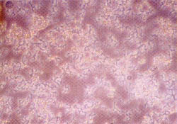
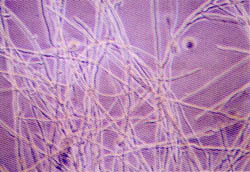
bacterial or fungal was more common with skin specimens taken from burned
patients despite careful preparation (Fig. 1, a&b). Cultured allogenic grafts
were found to pro- mote healing provided that the recipient area was well
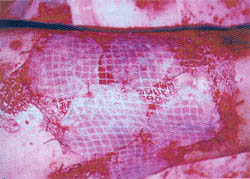
prepared. In case #1, complete closure of the interstices of the meshed
graft occurree in 5 days (Fig. 2, a&b) and full epithelializatio of the
STSG donor site was observed in 6 day (Fig. 2, a&b). Control areas in the
same pane required 9 and 11 days respectively for co plete healing. In the
second case, cultured all genic keratinocyte grafts reduced the areas full-thickness
burn by 70% in 5 days and pr moted healing of a rather deep STSG don site.
Graft loss occurred in case 3 due to imp fect immobilization and in case
4 due to infection.
|
|
| Fig. (1-A): Bacterial contamination
of keratincoyte culture. Inverted phase micrograph magnified
100 X |
Fig. (1-B): Fungal contamination
of keratinocyte culture. Inverted phase micrograph magnified
100 X. |
|
|
|
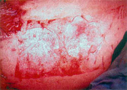
| Fig. (2-A) :Cultured allogenic keratinocyte
gafts applied onto meshed skin autograft. |
Fig. (2-B): The same case showing
complete closure of the interstices of the meshed autograft
by the 5th postoperative day. |
|
|
|
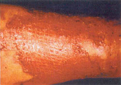
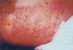
| Fig. (3-A) Split-thickness skin gaft
donor site covered by cultured allogenic keratinocyte grafts,
intraoperative view |
Fig. (3-B): The same donor site,
7 days postoperatively. See delayed healing of the periphery
in aereas not covered by cultured allograft. |
|
|
DISCUSSION
The clinical applications of tissue culture have been increasing since
the development of reproducible culture techniques. A recent method that
had been developed for wound closure is the use of cultured autologous keratinocyte
grafts that had been used to close burn wounds [4] and leg ulcers
[15].
Cultured autologous keratinocytes can permanently attach [5]. However,
they require the use of a biopsy from the skin of the patient and there
is a delay corresponding to the time required for cultivation of the epidermal
cells in vitro [6]. Another problem is the slow growth and reduced
culture life span for cells from old donors [16]. The epidermis produced
by cultured autologous keratinocyte grafts is unstable [17,18] and
5 years are needed for a neodermis to form [19]. Therefore, attention
was focused on the use of cultured autologous keratinocytes with dermal
equivalent [20,21]
. On the other hand, cultured allogenic keratinocyte grafts could be immediately
available, especially when cryopreserved without the need for skin biopsy
from the recipient [22,23]. It is also possible to growth cells from
young donors, which have a higher growth potential and better wound healing
power than cells from old cell donors [6,24]. Cultured allogenic
keratinocyte grafts were thought to take permanently because they do not
express the class II HAL-DR antigen [25,26]. However, further investigations
by several authors showed that cultured allogenic grafts undergo gradual
replacement by cells from the recipient without detectable rejection [27,28].
In our experimental work, we were able to prepare the MCDB-153 culture medium.
This medium was used to grow and propagate normal human adult keratinocytes
for 5 passages without the need of the special murine 3T3 feeder layer of
cells [14]. We also could promote differentiation and stratification
of PI and P2 cultures to prepare epidermal sheets in vitro suitable for
grafting. Contamination was responsible for culture failure during the in
vitro stage. This was common when the biopsy specimen was taken from burned
patients. The skin of these patients may be colonized by resistant bacteria
from the septic burn wounds, a problem that is not existing in non burned
patients and cases of elective aesthetic surgical procedures. This is why
we decided to start our tissue culture work by allogenic cells.
In this study, burn wounds and STSG donor sites were treated with cultured
allogenic keratinocyte grafts. Wound healing was accelerated as evidenced
by rapid closure of the interstices of the meshed STSG autograft (case 1),
prompt healing of residual deep burn wound (case 2) and early epithelialization
of STSG donor sites (cases 1 and 2). Promotion of healing would minimize
the period of hospital stay and allow reharvesting of the same STSG donor
sites in burned patients. Vol. 26, No. 2 / Cultured Allogenic Keratinocyte
Grafts
Considered as an indicator for promotion of burn wound healing rather than
permanent coverage. Cultured allogenic keratinocyte grafts probably exert
their effects on wound healing through the release of cytokines and growth
factors [29]. These factors act by stimulating the epidermal cells
from the margins of the raw area or from the meshed STSG in full-thickness
burn wounds and by stimulating dermal appendages in STSG donor sites and
partialthickness burn wounds. The attached allogenic cells are gradually
replaced by autologous cells from the recipient .without observable rejection
[27,28]. Failure occurred in two cases due to shearing and infection (cases
3 and 4).
We should emphasize that although successful attempts are reported in this
study, the areas treated by the cultured allografts represented only localized
parts of the expensive bum wounds. A similar observation was reported by
authors with cultured keratinocyte autografts [30,31): This was true especially
in case 2 in our study. The cost effectiveness is not studied for this research
procedure.
Summary:
This study represents the first Egyptian report on the in vitro cultivation
of normal human keratinocytes into stratified sheets suitable for grafting.
We also described the clinical application of these grafts to treat a limited
number of cases of burn wounds and STSG donor sites. The procedure requires
strict aseptic laboratory technique and good preparation of the cultured
graft recipient sites. The ability to grow normal. human keratinocytes in
vitro opens the door for laboratory research on skin cells, including work
on composite skin substitutes propriate dermal equivalents with the epidermal
cells.
REFERENCES
-
Madden MR, Finkelstein J.L., Staiano-Coico 1 Goodwin C.W., Shires G.T., Nolan E.E. and Heft J.M.: Grafting of cultured allogenic epidermis on s and and third degree burn wounds in 26 patients. Trauma., 26: 955-962, 1986.
-
Banks-Schlegel S. and Green H.: Formation of epid mis by serially cultivated human epidermal ce transplanted as an epithelium to athymic mice. Tr
plantation, 29: 308-313, 1980.
-
Eisinger M., Monden M., Raaf J.H. and Former J.1 Wound coverage by a sheet of epidermal cells gr43 in vitro from dispersed single cell preparations. ~Surgery, 88: 287-293, 1980.
-
O'Connor N.E., Mulliken J.B., Banks-Schlegel S., Kehinde O. and Green H.: Grafting of burns with cultured epithelium prepared from autologous epidermal cells. Lancet, 1: 75-78, 1981.
-
Gallico G.G., O'Connor N.E., Compton C.C., Kehinde O. and Green H.: Permanent coverage of large burn wounds with atuologous cultured human epithelium. N. Engl. J. Med., 311: 448-451, 1984.
-
Phillips T.J., Kehinde O., Green H. and Gilchrest B.A.: Treatment of skin ulcers with cultured epidermal allografts. J. Am. Acad. Dermatol., 21: 191-199, 1989.
-
El-Khatib H.A.: Skin substitutes for burns. M.D. Thesis. Supervised by: El-Falaky, M.H. and Hansbrough J.F., Al-Azhar University, 1989.
-
Rizk LM.: Early surgical excision and coverage of burns, a clinical and immunological study. M.D. Thesis. Supervised by: Talaat H.A.F., Kamal M.S. and Kamal K., Cairo University, 1991.
-
Ahmad A.H.A.: A study on cultivation of autologous epidermal cells and their use as grafts for coverage of skin ulcers and deep burn wounds. M.D. Thesis. Supervised by: Badran H.A., Khalifa A., Marcelo C.L., El-DeebT.M. and Mady E.A., Ain Shams University, 1997.
-
Marcelo C.L., Duel E.A., Rhodes L.M. and Dunham W.R.: An in vitro model of essential fatty acid deficiency. J. Invest Dermatol., 99: 771-777, 1992.
-
Boyce S.T. and Ham R.J.: Calcium-regulated differentiation of normal human epidermal keratinocytes in chemically defined clonal culture and serum-free serial culture. J. Invest. Dermatol., 81 Suppl (1): 33s-40s, 1983.
-
Boyce S.T. and Ham R.G.: Cultivation and frozen storage of normal epidermal keratinocytes in serumfree medium. J. Tiss. Cult. Meth., 9: 83-93, 1985.
-
Wille J.J., Pittelkow M.R., Shipley G.D. and Scott R.E.: Integrated control of growth and differentiation of normal human prokeratinocytes cultured in serumfree medium: Clonal analysis, growth kinetics and cell cycle studies. J. Cell. Physiol., 121: 31-44, 1984.
-
Green H., Kehinde O. and Thomas J.: Growth of cultured human epidermal cells into multiple epithelia suitable for grafting. Proc. Natl. Acad. Sci., Usa, 76: 5665-5669, 1979.
-
Hefton J.M., Madden M.R., Finkelstein J.L. and Shires G.T.: Grafting of burn patients with allografts of cultured epidermal cells. Lancet, 20: 428-430, 1983.
-
Barrandon Y. and Green H.: Three clonal types of keratinocyte with different capacities for multiplication. Proc. Natl. Acad. Sci., USA, 84: 230-2306, 1987.
-
Petersen M.J., Woodley D.T., Stricklin G.P. and O'Keefe E.J.: Production of procollagenase by cultured human keratinocytes. J. Biol. Chem., 262: 835840, 1987.
-
Woodley D.T., Peterson H.D., Herzog S.R., Strickin G.P., Burgeson R.E., Briggaman R.A., Cronce D.J. and O'Keefe E.J.: Burn wounds resurfaced by cultured autografts show abnormal reconstitution of anchoring fibrils. JAMA, 259: 2566-2571, 1988.
-
Compton C.C., Gill J.M., Bradford D.A., Regauer S., Gallico G.G. and O'Connor N.E.: Skin regeneration from cultured epithelial autografts on full thickness burn wounds from 6 days to 5 years after grafting. Laboratory Investigation, 60: 600-612, 1989.
-
Heck E.L., Bergestresser P.R. and Baxter C.R.: Composite skin gaff: Frozen dermal allografts support the engraftment and expansion of autologous epidermis. J. Trauma, 25: 106-112, 1985.
-
Hansbrough J.F., Boyce S.T., Cooper M.L. and Foreman T.J.: Burn wound closure with cultured autologous keratinocytes and fibroblasts attached to a collagen-glycosaminoglycan substrate. JAMA, 262: 21252130, 1989.
-
Teepe R.G.C., Koebrugge E.J., Ponec M. and Vermeer B.J.: Fresh versus cryopreserved cultured allografts for the treatment of chronic skin ulcers. Br. J. Dermatol., 122: 81-89, 1990.
-
Madden M.R., LaBruna A.A., Hajjar D.P. and Staiano-Cico L.: Transplantation of cryopreserved cultured epidermal allografts. J. Trauma, 40: 743-750, 1996.
-
Hansbrough J.F.: Wound coverage with biologic dressings and cultured skin substitutes. R.G. Landes Company. Austin, pp 21-114, 1992.
-
Morhenn V.B., Benike C.V., Cox A.J., Charron D.J. and Engleman E.G.: Cultured human epidermal cells do not synthesize HLA-DR. J. Invest. Dermatol., 78: 32-37, 1982.
-
Thivolet J., Faure M., Demiden A. and Mauduit G.: Long-term survival and immunological tolerance of human epidermal allografts produced in culture. Transplantation, 42: 274-280, 1986.
-
Gielen V., Faure M., Mauduit G. and Thivolet J.: Progressive replacement of human cultured epithelial allografts by recipient cells as evidenced by HLA class 1 antigen expression. Dermatologica., 175: 166-170, 1987.
-
Van der Merwe A.E., Matheyse F.J., Bedford M., Van Helden P.D. and Rossouw D.J.: Allografted keratinocytes used to accelerate the treatment of burn wounds are replaced by recipient cells. Burns, 16: 193-197, 1990.
-
Boyce S.T.: Epidermis as a secretory tissue. J. Invest. Dermatol., 102: 8-10, 1994.
-
Rue L.W., Cioffi W.G., McManus W.F. and Pruitt B.A.: Wound closure and outcome in extensively burned patients treated with cultured autologous keratinocytes. J. Trauma, 34: 662-667, 1993.
-
Sheridan R.L. and Tompkin D.J.: Recent clinical experience with cultured autologous epithelium. Br. J. Plast. Surg., 49: 72-73, 1996.
|